Magneto/electroencephalography (M/EEG) studies of dreaming are an essential paradigm in the investigation of neurocognitive processes of human consciousness during sleep, but they are limited by the number of observations that can be collected per study. Dream research also involves substantial methodological and conceptual variability, which poses problems for the integration of results. To address these issues, here we present the DREAM database—an expanding collection of standardized datasets on human sleep M/EEG combined with dream report data—with an initial release comprising 20 datasets, 505 participants, and 2643 awakenings. Each awakening consists, at minimum, of sleep M/EEG ( ≥ 20 s, ≥100 Hz, ≥2 electrodes) up to the time of waking and a standardized dream report classification of the subject’s experience during sleep. We observed that reports of conscious experiences can be predicted with objective features extracted from EEG recordings in both Rapid Eye Movement (REM) and non-REM (NREM) sleep. We also provide several examples of analyses, showcasing the database’s high potential in paving the way for new research questions at a scale beyond the capacity of any single research group.
A third of the life of a healthy adult is spent asleep and, for a portion of that, dreaming. Throughout the night, during any stage of sleep, subjective conscious experiences can occur repeatedly—what we broadly call dreams. Dream studies, producing combined sleep and dream data, support various ongoing topics of research, including clinical (e.g., parasomnias), neurocognitive (e.g., learning and memory), and basic ones (e.g., the neural correlates of consciousness in dreaming). In this work, we present the Dream EEG and Mentation (DREAM) database, which gathers and standardizes multicentric recordings of electroencephalography (EEG), magnetoencephalography (MEG), and dream reports to advance the aforementioned research topics.
Commonly, people think about dreams as being immersed in a lifelike world, disconnected from the external environment, in which perceptually rich and narrative experiences may be had1,2. However, sleep is accompanied by many types of conscious experiences that do not match this notion. In the transition from wake to sleep and vice versa, bizarre perceptual experiences may be reported, which are known as hypnagogic and hypnopompic hallucinations, respectively3,4,5,6. Minimal experiences of static, fragmentary, unimodal percepts or simple thoughts are often called sleep mentation. Some dreams are lucid: when dreamers are aware that they are dreaming and may be able to control the oneiric content7. Some other dreams include “false awakenings”, during which participants believe they have awoken but are actually still dreaming, sometimes giving the sensation of a dream within a dream8. A dream can be called a nightmare when it includes experiencing disturbing emotions, such as intense fear, anxiety, or grief, which may trigger an awakening. Frequently occurring nightmares can be a sign of an underlying mental or neurological disorder, such as post-traumatic stress disorder or narcolepsy9,10,11,12. In addition, the so-called “white dreams” are reported by awakened participants as a dream experience that they are convinced occurred but cannot recall the specific contents of13. Finally, there are periods of sleep in which experience is reported to have been absent altogether. These reports either signify unconsciousness during sleep or lack of any recall of dream experiences. Notably, the above delineations are not always unambiguously recorded in sleep studies, making data reuse between different research topics difficult. For this paper, we will broadly define dreams as any experiences recalled from sleep.
Subjective verbal reports provided upon awakening are considered the gold standard for collecting data from dream experiences in current research practices. Yet, the information we can extract from verbal reports has limits and tradeoffs. Sleep must be interrupted in order for participants to report; the quality of reports may be impaired due to confusion or sleepiness (e.g., sleep inertia), and the comprehensiveness of reports diminishes with time due to forgetting and the complex act of reporting14,15. As such, depending on the research question, many awakenings must be performed to produce reliable data. In the special and rare case of lucid dreaming, participants can produce rudimentary but objective signals without waking up, in the form of voluntary eye16, facial, or respiration17 movements, or fist clenching18,19,20.
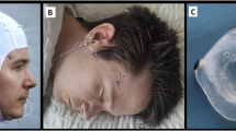
The classic stages of sleep were identified based on visual examination of the polysomnography (PSG), which includes the simultaneous recording of the electroencephalogram (EEG, for brain activity), electrooculogram (EOG, for eye movement), and electromyogram (EMG, for muscle activity)21,22,23. According to the American Academy of Sleep Medicine24, wakefulness features low-amplitude, high-frequency EEG activity, active EOG, and high EMG tone. When individuals keep their eyelids closed and relax, alpha rhythms (8−12 Hz) predominate. NREM stage 1 (N1 or sleep onset) presents a slowing of the EEG toward theta waves (4−7 Hz), slow rolling eye movements in the EOG, and reduced EMG activity. NREM stage 2 (N2 or light sleep) is marked by sleep spindles (a transient 12−16 Hz oscillation) and K-complexes in the EEG, no eye movements in the EOG, and further decreased EMG tone. NREM stage 3 (N3, deep sleep, or slow-wave sleep), shows trains of high-amplitude delta waves (0.5−4 Hz) in the EEG, absent EOG activity, and minimal EMG tone. REM sleep is identified by low-amplitude, mid-low to high frequencies EEG activity, rapid eye movements in the EOG, muscle atonia in the EMG, and has been traditionally linked to dreaming.
Early sleep studies utilizing PSG suggested that dreams occur mostly during a distinct stage of sleep, the REM sleep, and only rarely during non-REM (NREM) sleep25,26. However, many subsequent studies have revealed the connection between dreaming and sleep stages to be more complex (e.g.27,). It is now established that dream reports accompany about 85% of awakenings from REM sleep and about 40−60% from NREM sleep28,29,30, varying as the night progresses31,32. While dream reports from NREM sleep tend to be less frequent and have less vivid and detailed content compared to those obtained from REM sleep, NREM dreams occurring at sleep onset (N1) can resemble REM dreams in vividness and bizarreness (despite having a shorter duration)3,33, and morning dreams from both REM and NREM sleep may be indistinguishable from each other34. Generally, the frequency of dream recall in both NREM and REM sleep increases in the latter half of the night, and so does dream complexity35,36. Thus, REM sleep is neither necessary nor sufficient for dream experiences37; dreams can occur in neurophysiologically distinct stages of sleep.
Objective and consistent markers of dreaming, or dream recall, are still being investigated. Recent studies suggest that the occurrence of reported dreams has a spectral EEG marker consistent between and within sleep stages: a reduction of low frequency delta power38,39,40,41,42,43,44,45,46. Other frequency bands have been reported to be associated with certain aspects of dreams47,48,49,50,51,52,53,54,55, and so have other features of EEG such as spindles or network properties56,57,58,59. In addition, different aspects of neural correlates of dreaming may be revealed by using MEG in addition to EEG because of their methodological differences. EEG studies investigating the neural correlates of specific contents of dreaming42,60,61,62,63,64, revealed a homogeneity of the neural basis of subjective experiences between wakefulness and sleep. Similarly to the waking brain, affective processing in dreams is associated with frontal alpha asymmetry63, dreams involving kinesthesia and movement are linked to inter-hemispheric coherence of beta oscillations64, perceptual contents of dreams correlate with gamma activity in the occipital, temporal, and parietal regions, and thought-like mentation is associated with frontal activation in the gamma frequency range42. However, more research is needed to determine exact associations between multidimensional EEG patterns, type of conscious experience and specific dream contents.
Science has not yet determined whether or not awareness is present in cases where behavioral reports are unavailable. This problem is most prevalent in everyday sleep, but the implications run deeper when considering other conditions involving unresponsiveness. We can have no ease of certainty about the absence of consciousness in patients diagnosed with unresponsive wakefulness syndrome65, or the depth of general anesthesia needed to prevent individual intraoperative awareness66. Indeed, phenomenologically, such intraoperative awareness is often reported/described as intraoperative dreaming67. Given these phenomenological similarities, it is hypothesized that discovering the neural correlates of consciousness in dreaming would help solve these problems in detecting consciousness in the absence of reports. Combined sleep and dream data provide a promising paradigm for such research.
Unfortunately, sleep studies that include dream sampling protocols require a huge amount of time, skills, and resources. Only a few laboratories can afford to perform this kind of investigation. Moreover, such studies often have to rely on relatively small sample sizes. Indeed, previous neural correlates studies of dreaming have usually been performed on sample sizes in the order of tens of awakenings, thus leading to reservations about the robustness and generalization of findings68 and slowing the pace of progress. Studies collecting hundreds of awakenings are rare (e.g., Refs. 42,44). As such, there is a significant communal benefit to having a large database of existing dream and EEG datasets contributed by the research community available. Such collaborative efforts are multiplying in the field of neuroscience (e.g., Allen Institute for Brain Science, Stanford Center for Reproducible Neuroscience, Refs. 69,70), but until the current project, there has been no corresponding endeavor specifically for neurobiological dream research. Instead, existing related databases offer either sleep EEG data without dream reports (e.g., the National Sleep Research Resource71) or dream report data without neurophysiological recordings (e.g., DreamBank.net72 and the Sleep and Dream Database https://sleepanddreamdatabase.org/).
A major challenge that must be overcome to build a reuse-friendly database is ensuring that all datasets, which originate from primarily unrelated studies, are comparable. We, therefore, propose a unified dream classification system that delineates the types of dreams relevant to consciousness research to provide unambiguous and comparable categorizations between studies. We also implement a consistent, high standard for data formatting and documentation. Pertinent metadata are extracted from each datum in the collection and entered into the database for search and browsing.
The present work describes the Dream EEG and Mentation (DREAM) database, created to facilitate and stimulate dream research and collaborative studies using a neuroscientific approach. The DREAM database is released with an initial 20 data sets, composed of 505 participants and 2643 awakenings. Each datum represents PSG data of a sleep period leading up to awakening and a subjective report of the sleep experience. The database is openly accessible from https://monash.edu/dream-database. Furthermore, the database is open to new data contributions and will continue to expand indefinitely. It is maintained by a core team of scientists from different institutions on a voluntary basis.
Results
In the following section, we demonstrate the use of our database by analyzing behavioral, experiential, and electrophysiological data. For a detailed description of the methods see Supplementary Methods 2. For this analysis, we checked critical metadata and selected relevant datasets using the database without performing a time-consuming download of huge amounts of data, demonstrating the usefulness of the dataset. From the DREAM database, we retrieved relevant data, such as the dream report classification and sleep stage of the pre-report epoch. We also retrieved the age and sex of the participant if provided.
Behavioral analyses
Using combined dream report classifications and manually scored sleep stages of the final epoch, provided by contributors as aggregated in the DREAM database Data records table (version 1), we performed statistical analyses for the relationship between sleep stage and dream experience. Based on the previous literature, we expected the relative frequency of sleep experience categories to be different between awakenings from different sleep stages (e.g., Nielsen, 200028). Qualitatively, we expected deeper NREM sleep to be associated with less frequent reports of dreaming and REM sleep to be associated with more frequent reports of dreaming than NREM sleep.
We observed a dependence between the Last sleep stage and Experience (Table 4) with high statistical significance (chi-squared test, X2 = 120.9, df = 6, p < 10-15). We also observed the expected qualitative trend of deeper NREM sleep stages associated with fewer reports of “Experience”, and an association between NREM sleep depth with reports of “No experience” and reports of “Experience without recall”. The REM sleep stage was associated with fewer “No experience” and “Experience without recall” reports and more “Experience” reports than expected with the independence assumption (Supplementary Table 4).
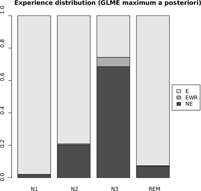
The behavioral data were further analyzed using generalized linear mixed-effects modeling to account for the grouping effects of the experimental study and individual participants. See Supplementary Methods 2 for method’s details and Supplementary Table 5 for the estimated fixed effects coefficients, which are visualized in Fig. 1.
Fig. 1: Probability of reports per sleep stage.
Y-axis shows the maximum a posteriori probability for the three types of reports (E: “Experience”, EWR: “Experience without recall”, NE: “No experience”), split by each sleep stage (x-axis).
Based on the fitted model’s posterior distribution, the following inferences could be made with high confidence (>99% density in the region of interest). We found the expected qualitative trend of deeper NREM sleep stages associated with lower odds of “Experience” against “No experience” reports. There was no clear trend for the odds of “Experiences without recall” against “No experience” reports with respect to NREM depth. The odds of “Experience” against “No experience” reports for the REM sleep stage were higher than for N2 and lower than for N1.
EEG analyses
We analyzed EEG recordings of selected DREAM datasets in two respects: power spectral density and automated sleep scoring. Of the datasets available at the time of the DREAM database’s first version, we selected six datasets suitable for our EEG analyses: Oudiette N1Data, Zhang & Wamsley 2019, De Gennaro Multiple Awakenings, De Gennaro Young Adults, Tononi Serial Awakenings, and REM Turku (see Table 3 for details). For brevity, in our results, they are named following the corresponding article’s first author: Lacaux, Zhang, Scarpelli 1, Scarpelli 2, Siclari, and Sikka, respectively. The rest of the datasets were excluded because of the use of nonstandard equipment (e.g., dry EEG headsets), the presence of stimuli during sleep, or the participants’ demographics (e.g., younger or older participants). We focused on the 30 s of sleep before each dream report and used the Records.csv tables from each dataset to assign those epochs to their respective sleep stages and dream experiences. The three primary categories of dream experience were included—i.e., “Experience”, “Experience without recall” and “No experience”—comprising a total of 1462 30-s epochs prior to a report. We performed minimal preprocessing where appropriate.
Power spectral densities
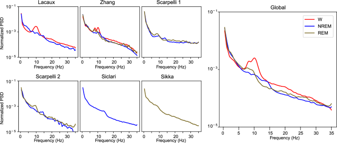
We computed the power spectral densities (PSDs) associated with Wake (W), NREM, and REM sleep stages from one central electrode (C3 or C4). We found the stereotypical alpha peak (8–12 Hz) in W, the sigma (spindle) peak (12–15 Hz) in NREM, and an increase in the theta range (4–8 Hz) in REM (Fig. 2).
Fig. 2: Sleep stage power spectral densities.
Left (smaller) panels show the average normalized PSD for the 30-s epochs prior to a report for each database. Not all databases have all the sleep stages. The right panel shows the average of 1462 epochs from the 6 analyzed databases. Colors denote different sleep stages.
Automatic sleep scoring
We applied a previously validated automatic sleep scoring algorithm73 to the six datasets to estimate hypnodensities, which are probability distributions of the scored sleep stage for a given epoch. As the algorithm operates on 15-s epochs rather than 30, we averaged hypnodensities over the last two 15-s epochs prior to a dream report. For this analysis, we used two EEG channels (a central and occipital channel), two EOG channels (left and right), and one chin EMG channel.
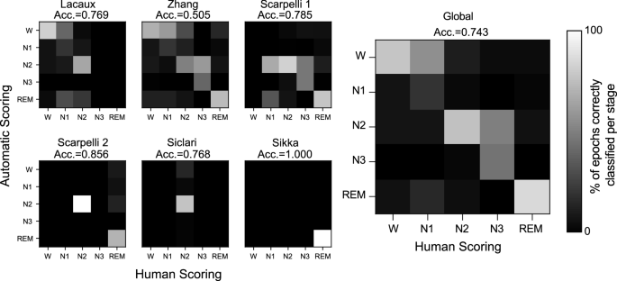
Results are given in Fig. 3 as confusion matrices against human scoring. The sleep scoring algorithm yielded an average accuracy above .72, which was close, despite the lack of context, to the inter-human scoring accuracy following the American Academy for Sleep Medicine (AASM) guidelines22.
Fig. 3: Automatic sleep scoring.
The left panels show the confusion matrices for each database. Under each database name, the average subset accuracy score is shown (see Supplementary Methods 2 for details). The right panel shows the confusion matrix when all 1,462 epochs are considered, along with their respective average subset accuracy scores.
Combined dream reports and EEG analysis
Prediction of experience from EEG activity
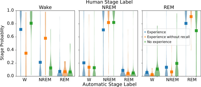
New automated algorithms to score sleep EEG recordings offer a probabilistic, rather than discrete, approach to sleep staging73,74. This probabilistic approach can reveal subtler differences that could be masked by the classical categorical view of sleep stages. We thus examined whether the dream report category was reflected in the probability of each sleep stage (hypnodensities, Fig. 4). We performed a Bayesian ANOVA on the hypnodensities, modeling the main effects of automatically-scored sleep stage and dream report and a random grouping effect for each dataset. Note that we reduced three NREM categories (N1, N2, and N3) into a single factor of NREM for this analysis. Overall, hypnodensities tracked human sleep scoring (i.e., the stage with the highest probability in the hypnodensity was congruent with human-scored sleep stages) with a significant main effect of sleep stage on hypnodensities (Bayes factor, log(BFinclusion) = +∞). Hypnodensities were also modulated by the dream report category, with both a main effect of report category (Bayes factor, log(BFinclusion) = 8.582) and interaction between categories and sleep stages (Bayes factor, log(BFinclusion) = 10.367). Post-hoc estimates indicate that NREM epochs associated with dream experience show an increased probability mass toward W (non-overlapping 95% credible intervals; Bayes factor, log(BFmodel) = 96.7).
Fig. 4: Hypnodensities for each reduced human-scored sleep stage.
Distributions of hypnodensities per reduced sleep stage (where NREM = N1 + N2 + N3) for 1,462 pre-report epochs. All epochs belonging to the same sleep stage and dream experience were pooled together. Colors denote different categories of dream experience and squares the distribution average.
Combined dream reports and EEG analysis
Finally, we quantified the amount of information needed to successfully classify dream experience (“Experience” or “No Experience”) from EEG activity both in NREM and REM sleep. For this analysis, we used only three electrodes (F4, C4, and O2) that were commonly used in the six aforementioned studies. We extracted traditional PSD features and non-traditional features obtained from the catch22 analytical pipeline75 (see Supplementary Methods 2 for details). Catch22 provides 22 minimally redundant features that capture complex and nonlinear characteristics of time series. Only NREM (N1 + N2 + N3) and REM 30 s epochs prior to a report were considered in further analysis. NREM consisted of 778 epochs with 252 NE epochs and 456 E epochs. REM consisted of 342 epochs with 51 NE epochs and 282 E epochs. We computed three sets of features for each electrode:
1.
PSD: Normalized spectral power on 6 different frequency bands: delta (0.5–4 Hz), theta (4.1–8), alpha (8.1–11 Hz), sigma (11.1–15 Hz), beta (15.1–20 Hz) and gamma (20.1–35 Hz). In total, 18 features.
2.
Catch22bb: catch22 features computed for the broadband (bb) EEG signal (0.5–35 Hz). In total, 66 features.
3.
Catch22bf: catch22 features computed for the EEG signal band-filtered (bf) on the 6 aforementioned frequency bands. In total, 396 features.
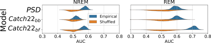
We found that for both NREM and REM “Experience” could be significantly discriminated (Bonferroni corrected p < 0.001, Wilcoxon rank sum test vs null distribution) from “No experience” using both PSD and catch22 features (Fig. 5). For NREM, the highest performance was obtained for PSD features (average AUC = 0.586, 5-th percentile = 0.505, 95th percentile = 0.608), while for REM the highest performance was obtained for Catch22bf (average AUC = 0.700, 5-th percentile = 0.663, 95-th percentile = 0.731). These results demonstrate that the DREAM database can be exploited to effectively investigate the neural correlates of dreaming. Furthermore, our results suggest that a more complex and nonlinear analysis –beyond PSD– may provide information relevant to decoding dreaming experiences from EEG recordings.
Fig. 5: Classification accuracy of dreaming experience (Experience vs. No experience) using EEG features.
Cross validation performance (in terms of area under the receiver operator characteristic curve, AUC, x-axis) from classifiers. Each of six classifiers was separately trained to discriminate dream experience between “Experience” and “No Experience” within NREM (left) and within REM (right) sleep, using a distinct set of features. Blue distributions (empirical) are the AUC values for test data from 200 random train-test splits of the data, while the orange ones (shuffled) are the AUC values for test data obtained by shuffling the labels for dreaming, serving as the null distribution. All empirical AUC distributions were significantly different from their respective null distributions (two-sided Bonferroni corrected Wilcoxon rank sum p-value < 0.001).
Discussion
Many research areas share a need for, yet lack of, easily-accessible simultaneous sleep and dream data. One of our primary intentions for creating the DREAM database is to satisfy this need in the area of dream research. The DREAM database will: (1) enhance comparability across studies with an agreed dream report classification scheme (Tables 1 and 2), using combined sleep and dream data, (2) allow the data to be easily found and reused, and (3) help mitigate the problems related to small datasets and lack of statistical power. With the DREAM database, we present a variety of combined sleep and dream data sets in a novel, standardized, human- and machine-readable format, as recommended by the FAIR guiding principles76. Detailed metadata of all datasets are kept by the DREAM database proper and made openly accessible. Furthermore, we have been actively curating and expanding the database with new dataset submissions from community contributors and will continue to do so indefinitely.
The DREAM database is designed to enable researchers to more easily analyze data on automated end-to-end analysis pipelines as we demonstrated (Figs. 3–5). The database offers a great variety of recordings and includes dream reports collected both from REM and NREM sleep. Combined data unambiguously confirmed that dreaming prevalence decreases with sleep depth in NREM (Tables 3 and 4, Fig. 1). We also show that dreams in NREM sleep are associated with a higher probability of wake-like activity, suggesting that dreams could reflect a phenomenon of ‘covert wake’ akin to the covert REM hypothesis proposed previously28 for NREM dreams (Figs. 3 and 4). Finally, we show that experience-relevant information can be decoded from EEG analysis both in REM and NREM sleep (Fig. 5).
The two latter findings should be further investigated in future studies, which goes beyond the scope of this paper. Our primary aim here is to present the database and demonstrate its use and potential. While useful on its own, the database and preliminary results will serve as the most promising starting point for future studies, to be published as the registered report or registered analysis format77, which came about in response to the replicability crisis78,79,80,81. Pre-registered studies require the authors to precisely specify their methods in advance of their study’s commencement, thus minimizing the risk of potential abuse of experimental and statistical methods—the so-called “data dredging” or “p-hacking”. This database could inform sample sizes and narrow the hypotheses’ space for new studies.
Participating journals are also required to make their decision to publish a registered study—based on the merit of the study’s methods, before the study can commence and regardless of its eventual results—thus reducing the file drawer effect (i.e., not publishing null results that can potentially contradict previous findings). As combined sleep and dream data is relatively expensive to produce, increasing its sharing can also help boost the number of replications and novel analyses that can be performed, accelerating scientific progress.
Sleep research is attracting attention as it can host new experimental paradigms for investigating the neural correlates of consciousness, like the no-report within-state approach82. Dreams represent instances of human experience that largely have no direct sensory input from, nor observable behavioral output to, the external world. Thus, the observed neural processes for conscious experience are much less attributable to confounding factors such as preconscious sensory processing or task-related activity82,83,84,85,86,87. By contrasting dreamless, unconscious sleep with dreamful, conscious sleep, the distinction between levels of consciousness within the same brain state becomes possible.
As the database continues to grow, we hope it can help answer some of the remaining basic questions about sleep and dreams. Why are we sometimes conscious and sometimes unconscious during sleep? Why do some people remember their dreams every day while others have very low dream recall frequency? Why do so few individuals regularly have lucid dreams, while others can realize they are dreaming and control their oneiric content? Ultimately, discovering the neural correlates of specific contents of dreaming, which may require substantially more data than the classification of the presence or absence of dreaming experience, will play a significant role in understanding the neural mechanisms of consciousness. We look forward to seeing how future studies utilize the datasets presented here to tackle these and other questions and possibly identify new lines of research.
Methods
Ethics
The Dream EEG and Mentation database project was approved by the Monash University Human Research Ethics Committee (Project ID 31269). Furthermore, the authors of the originating studies of DREAM datasets confirm that ethical approval for their respective studies were obtained from the relevant ethical committees, that participants provided informed consent and consented to sharing and secondary use of the data. The corresponding authors of these and future studies included by the database are obliged to protect participants’ rights by one or a combination of: obtaining the participants’ consent, ensuring the dataset is published with the appropriate accessibility, redacting data, and irreversibly de-identifying the data.
Dataset selection criteria
The DREAM database has an ongoing call for data consisting of PSG of human participants prior to waking and associated reports of conscious experiences (or lack of such) of that sleep period. The selection criteria were arrived at by consensus among the researchers who answered our first call for participation.
The recommended minimal technical standards are:
11.
At least two referenced EEG electrodes localizable by the 10-5 system;
12.
At least 20 s of continuous sleep recorded up until the moment of awakening;
13.
At least 100 Hz EEG sampling rate;
14.
Raw data or minimally preprocessed data.
We also ask data contributors to include the following information.
21.
Dream report classification for each datum (see Table 1) and the original dream report or data concerning it if possible;
Table 1 Dream report classifications
22.
Dream report categorization protocol, if nonstandard (see Table 2);
Table 2 Dream categorization rules and examples of their application
23.
De-identified participant labels for each datum;
24.
Treatment/condition/experimental group labels for each datum, if any;
25.
Any notable observations or artifacts;
26.
A description of the study protocols in enough detail to allow the replication of experimental procedures;
27.
Special instructions for decoding data files, if any.
Dream reports may include oral report transcriptions, written reports by study participants, interview responses, and inventory scores, but the minimum requirement is that the reports are categorized, where possible, into one of the following three classifications by the contributor: “experience”, “experience without recall”, and “no experience” or otherwise in combined classifications (where data are ambiguous). We describe the three main classifications in Table 1 (see below).
Data contributors are required to make their datasets available, either fully open-access or mediated-access, depending on what was approved by their own ethics committee.
Dream report categorization
We have defined dreaming to have a very broad meaning here, both for simplicity and for inclusivity with regard to the database. Our definition of dreaming includes all forms of conscious experience occurring within any stage of sleep that the participant can recall, regardless of their specific contents. It does not distinguish between immersive, multisensory, emotional and narratively complex experiences and experiences involving only thinking, mentation, or isolated forms of imagery—for example, visual or auditory. Our database uses a unified system for classifying dream reports into one, or a combination, of three ordinal categories: “experience”, “experience without recall” (also known as “white dreams”), and “no experience.” Their definitions are given in Table 1. As the contributed datasets can include additional, detailed subjective reports, it is possible to reclassify a subset of the datasets using a different set of definitions to the extent that the information is made available.
Since not all datasets may have originally collected and categorized data according to DREAM’s definitions, the standardized dream report classification may have been recategorized from an original categorization or raw data. The procedures used for obtaining these are given in the dataset’s description if applicable. Specifically, we applied the following rules (see also Table 2).
1.
If original categories are analogous or fit into the standard definitions, then a direct mapping of the categories is acceptable.
2.
If original categories fit into none or more than one of the standard definitions—but the raw reports contain data that allow for reclassification—then a reclassification is provided in addition to the original classification.
3.
If original categories do not fit into the standard definitions unambiguously—and the raw reports do not contain data that allow for reclassification—then the original classification is provided and its categories are interpreted as the combination of all standard definitions that could apply to it.
Data postprocessing
All datasets protect their subject’s confidentiality by redacting or removing names, dates of birth, addresses or other personally identifiable information.
PSGs including annotations are formatted as European Data Format “plus” (EDF + ) files. No further processing is applied to the signal post digitization unless otherwise specified in the study description, and any relevant processing of PSGs during their collection are noted.
Data Records
2643 data were collected in 20 separate studies. Descriptions of the individual datasets are given in Table 3. The registry of all datasets and their metadata constitute the main database and are searchable and downloadable at https://monash.edu/dream-database open access. Dataset packages are separately available either by open access or by request from the data contributor.
Table 3 The first set of datasets presented by the DREAM database
The DREAM database continues to be open to new contributions. Potential data contributors may visit the main website to receive instructions on how to format and upload their datasets.
Dataset format
Each dataset is packaged in a standardized file directory structure convention. PSG data are found in the “Data/PSG” subdirectory, dream report data are found in the “Data/Reports” subdirectory, and other miscellaneous data are found under the “Data” directory. Metadata for each awakening (see Supplementary Table 1) and the study’s experimental description are found in the root directory.
PSG data are presented in European Data Format (EDF)–compatible files, where each file contains data prior to a report. The recording length of the EDF file depends on the study design and is specified for each dataset. The Subject ID and Case ID values for each datum are encoded in the EDF header, specifically in the local patient identification field, whose format is analogous to the EDF+ standards. The Subject ID is given in the patient code subfield, and the Case ID is given in the patient name subfield.
Database format
The database records metadata of each awakening from every study in the table “Data records” as a single record with 20 fields as listed in Supplementary Table 2. Additionally, metadata of each unique study of origin are recorded in the table “Datasets” as a single record with 31 fields listed in Supplementary Table 3.
These records can be downloaded as whole, are open access and searchable, allowing researchers to discover data relevant to their work easily. The database is updated when new datasets are added, or amendments are made.
Technical validation
PSG artifacts were detected via visual inspection and noted by data contributors. From this, data that were found to contain =['<10 s of artifact-free EEG in the final 20 s were excluded from the datasets.
Database curators checked datasets via a combination of manual and automated checks for adherence to the DREAM dataset package standard. This included proper data quality (see Supplementary Methods 1), file directory structure, data file formatting, and metadata consistency.
Note also that manual sleep scoring can label a given epoch as Wake even before the participants were awoken (e.g., in serial awakening paradigms). In fact, there is abundant evidence for subjective-objective sleep discrepancies88. This happens in the context of our data, where either manual or automated EEG-based (objective) sleep stage classification labels an epoch as Wake, but during the same epoch, the participant reports to be asleep. For completeness, we have also included these subjective-objective discrepant epochs, as they may hold relevant EEG activity for dream experience. However, special attention should be paid to these epochs.
Table 4 Sleep stage vs. Experience contingency table
The DREAM database can be found from the landing page at https://monash.edu/dream-database or accessed directly at https://doi.org/10.26180/22133105. All DREAM datasets with open accessibility may be freely downloaded from the internet at their respective URL links recorded in the “Data URL” field of the “Datasets” table of the database (https://bridges.monash.edu/articles/dataset/The_DREAM_database/22133105?file=49774971). A detailed explanation of the columns of this table can be found on Supplementary Table S3. For datasets with more restricted access, a request for access may be made of the dataset’s corresponding author; details as to the extent of the restrictions, whether there are individual sharing agreements and a time frame of response to requests are given in the respective “Data restriction note” field of the “Datasets” table. Corresponding authors are recorded as uniquely identifying keys in the respective “Corresponding contributor ID” fields; the keys can be looked up in the “Key ID” field of the “People” table of the database, where you can find their contact information. A description for each dataset in the DREAM database at the time of writing is given below, in Table 3.
Sodré, M. E., Wießner, I., Irfan, M., Schenck, C. H. & Mota-Rolim, S. A. Awake or sleeping? Maybe both… a review of sleep-related dissociative states. J. Clin. Med.12, 3876 (2023).
Siclari, F., Valli, K. & Arnulf, I. Dreams and nightmares in healthy adults and in patients with sleep and neurological disorders. Lancet Neurol.19, 849–859 (2020).
HEARNE, K. M. T. Lucid Dreams: An Electro-Physiological and Psychological Study. (Thesis Ph. D., Liverpool University, 1978).
Dresler, M. et al. Neural correlates of dream lucidity obtained from contrasting lucid versus non-lucid REM. Sleep A Combined EEG/fMRI Case Study Sleep.35, 1017–1020 (2012).
Rechtschaffen, A. & Kales, A. A manual of standardized terminology, techniques and scoring system for sleep stages of human subjects. Arch. Gen. Psychiatry20, 246−247 (1969).
Silber, M. H. et al. The visual scoring of sleep in adults. J. Clin. Sleep Med.3, 121–131 (2007).
Berry, R. B. et al. Rules for scoring respiratory events in sleep: update of the 2007 AASM manual for the scoring of sleep and associated events. Deliberations of the sleep apnea definitions task force of the american academy of sleep medicine. J. Clin. Sleep Med.8, 597–619 (2012).
Dement, W. & Kleitman, N. The relation of eye movements during sleep to dream activity: an objective method for the study of dreaming. J. Exp. Psychol.53, 339–346 (1957).
Nielsen, T. A. A review of mentation in REM and NREM sleep: ‘covert’ REM sleep as a possible reconciliation of two opposing models. Behav. Brain Sc.i23, 851–866 (2000).
Noreika, V., Valli, K., Lahtela, H. & Revonsuo, A. Early-night serial awakenings as a new paradigm for studies on NREM dreaming. Int. J.Psychophysiol.74, 14–18 (2009).
Siclari, F., Larocque, J. J., Postle, B. R. & Tononi, G. Assessing sleep consciousness within subjects using a serial awakening paradigm. Front. Psychol.4, 542 (2013).
Cicogna, P., Natale, V., Occhionero, M. & Bosinelli, M. A comparison of mental activity during sleep onset and morning awakening. Sleep21, 462–470 (1998).
Casagrande, M., Violani, C., Lucidi, F., Buttinelli, E. & Bertini, M. Variations in sleep mentation as a function of time of night. Int. J. Neurosci.85, 19–30 (1996).
Moffitt, A. et al. Individual differences among pre- and post-awakening EEG correlates of dream reports following arousals from different stages of sleep. Psychiatric J. Univ. Ottawa7, 111–125 (1982).
Morel, C. R., Hoffmann, R. F. & Moffitt, A. R. The electrophysiological correlates of dream recall and nonrecall from stage 2 sleep. Can. J. Psychol. / Revue canadienne de psychologie45, 140–147 (1991).
Esposito, M. J., Nielsen, T. A. & Paquette, T. Reduced Alpha power associated with the recall of mentation from Stage 2 and Stage REM sleep. Psychophysiology41, 288–297 (2004).
Chellappa, S. L., Frey, S., Knoblauch, V. & Cajochen, C. Cortical activation patterns herald successful dream recall after NREM and REM sleep. Biol. Psychol.87, 251–256 (2011).
Siclari, F., Bernardi, G., Cataldi, J. & Tononi, G. Dreaming in NREM sleep: a high-density EEG study of slow waves and spindles. J. Neurosci.38, 9175–9185 (2018).
Scarpelli, S. et al. Electrophysiological correlates of dream recall during REM sleep: evidence from multiple awakenings and within-subjects design. Nat. Sci. Sleep12, 1043–1052 (2020).
Takeuchi, T., Ogilvie, R. D., Murphy, T. I. & Ferrelli, A. V. EEG activities during elicited sleep onset REM and NREM periods reflect different mechanisms of dream generation. Electroencephalograms. Rapid eye movement. Clin. Neurophysiol.114, 210–220 (2003).
Voss, U., Holzmann, R., Tuin, I. & Hobson, J. A. Lucid dreaming: a state of consciousness with features of both waking and non-lucid dreaming. Sleep32, 1191–1200 (2009).
Marzano, C. et al. Recalling and forgetting dreams: theta and alpha oscillations during sleep predict subsequent dream recall. J. Neurosci.31, 6674–6683 (2011).
Eichenlaub, J.-B. et al. Incorporation of recent waking-life experiences in dreams correlates with frontal theta activity in REM sleep. Soc. Cogn. Affect. Neurosci.13, 637–647 (2018).
Scarpelli, S. et al. Oscillatory EEG activity during REM sleep in elderly people predicts subsequent dream recall after awakenings. Front. Neurol.10, 985 (2019).
Scarpelli, S. et al. Dream recall upon awakening from non-rapid eye movement sleep in older adults: electrophysiological pattern and qualitative features. Brain Sci.10, 343 (2020).
Tagliazucchi, E. et al. Large-scale brain functional modularity is reflected in slow electroencephalographic rhythms across the human non-rapid eye movement sleep cycle. Neuroimage70, 327–339 (2013).
Eichenlaub, J.-B., Bertrand, O., Morlet, D. & Ruby, P. Brain reactivity differentiates subjects with high and low dream recall frequencies during both sleep and wakefulness. Cerebral Cortex24, 1206–1215 (2014).
Nielsen, T. et al. NREM sleep spindles are associated with dream recall. Res. Article1, 1 (2017).
Vallat, R. et al. Increased evoked potentials to arousing auditory stimuli during sleep: implication for the understanding of dream recall. Front. Hum. Neurosci.11, 132 (2017).
Sikka, P., Revonsuo, A., Noreika, V. & Valli, K. EEG Frontal alpha asymmetry and dream affect: alpha oscillations over the right frontal cortex during REM sleep and presleep wakefulness predict anger in rem sleep dreams. J. Neurosci.39, 4775–4784 (2019).
Mashour, G. A., Orser, B. A., Avidan, M. S. & Warner, D. S. Intraoperative awareness: from neurobiology to clinical practice. Anesthesiology114, 1218–1233 (2011).
Avidan, M. S. et al. Protocol for the BAG-RECALL clinical trial: a prospective, multi-center, randomized, controlled trial to determine whether a bispectral index-guided protocol is superior to an anesthesia gas-guided protocol in reducing intraoperative awareness with explicit recall in high risk surgical patients. BMC Anesthesiol.9, 8 (2009).
Wong, W. et al. The dream catcher experiment: blinded analyses failed to detect markers of dreaming consciousness in EEG spectral power. Neurosci. Conscious2020, niaa006 (2020).
Chambers, C. D. et al. Instead of “playing the game” it is time to change the rules: registered reports at <em>AIMS neuroscience < /em> and beyond. AIMSN1, 4–17 (2014).
Vul, E., Harris, C., Winkielman, P. & Pashler, H. Puzzlingly high correlations in fmri studies of emotion, personality, and social cognition. Perspect Psychol. Sci.4, 274–290 (2009).
Kriegeskorte, N., Simmons, W. K., Bellgowan, P. S. F. & Baker, C. I. Circular analysis in systems neuroscience: the dangers of double dipping. Nat. Neurosci.12, 535–540 (2009).
WW and NT were supported by National Health Medical Research Council (APP1183280) and by Japan Society for the Promotion of Science, Grant-in-Aid for Transformative Research Areas (N.T. 23H04829, 23H04830). JW, TA, and NT were supported by the Australian Research Council (NT DP240102680). G.B, V.E, D.B, M.B and G.A. were supported by the BIAL Foundation Grant Number #091/2020.
Funding
Open access funding provided by University of Skövde.
Author information
Author notes
These authors contributed equally: William Wong, Rubén Herzog.
These authors jointly supervised this work: William Wong, Rubén Herzog, Thomas Andrillon, Nicolas Decat, Valdas Noreika, Katja Valli, Jennifer M. Windt, and Naotsugu Tsuchiya.
Authors and Affiliations
School of Psychological Sciences and Turner Institute for Brain and Mental Health, Monash University, Clayton, Australia
William Wong & Naotsugu Tsuchiya
Institut du Cerveau - Paris Brain Institute - ICM, Inserm, CNRS, APHP, Hôpital de la Pitié Salpêtrière, Sorbonne Université, Paris, France
Rubén Herzog, Thomas Andrillon, Isabelle Arnulf, Nicolas Decat, Célia Lacaux & Delphine Oudiette
Brain Institute and Onofre Lopes University Hospital, Federal University of Rio Grande do Norte, Natal, Brazil
Department of Psychology, The University of Texas at Austin, Austin, USA
Benjamin Baird
School of Physiology, Pharmacology and Neuroscience, University of Bristol, Bristol, UK
Michele Bellesi
School of Biosciences and Veterinary Medicine, University of Camerino, Camerino, MC, Italy
Michele Bellesi
Department of General Psychology, University of Padova, Padova, Italy
Damiana Bergamo
Sleep Laboratory, School of Psychology, Swansea University, Swansea, UK
Mark Blagrove
Univ. Grenoble Alpes, Univ. Savoie Mont Blanc, CNRS, LPNC, Grenoble, France; Institut Universitaire de France (IUF), Paris, France
Jean-Baptiste Eichenlaub
Institute of Medical Psychology and Behavioral Neurobiology, University of Tübingen, Tübingen, Germany
Steffen Gais
Department of Psychology, University of Rome Sapienza, Rome, Italy
Luigi De Gennaro
Faculty of Design, Kyushu University, Fukuoka, Japan
Chihiro Hiramatsu & Yuki Motomura
Section for Physiology, MolMed Dpt., Institute of Basal Medical Sciences, Faculty of Medicine, University of Oslo, Oslo, Norway
Bjørn Erik Juel, Andre Sevenius Nilsen & Johan Frederik Storm
Center for Sleep and Consciousness, University of Wisconsin–, Madison, WI, USA
Bjørn Erik Juel
Department of Psychology and Cognitive Neuroscience Program, Northwestern University, Evanston, IL, USA
Karen R. Konkoly, Remington Mallett & Ken A. Paller
Institute of Psychology, Neuropsychology, University of Freiburg, Freiburg, Germany
Deniz Kumral, Jessica Palmieri & Monika Schönauer
Department of Neurology, Medical College of Wisconsin, Milwaukee, WI, USA
Joshua J. LaRocque
Association for independent research, Zurich, Switzerland
Bigna Lenggenhager
Centre for Brain and Behaviour, Department of Psychology, School of Biological and Behavioural Sciences, Queen Mary University of London, London, UK
Valdas Noreika
University of Geneva, Geneva, Switzerland
Lampros Perogamvros
Department of Psychology and Speech-Language Pathology, and Turku Brain and Mind Center, University of Turku, Turku, Finland
Antti Revonsuo, Pilleriin Sikka & Katja Valli
Department of Cognitive Neuroscience and Philosophy, University of Skövde, Skövde, Sweden
Antti Revonsuo, Pilleriin Sikka & Katja Valli
Department of Psychology, Swansea University, Swansea, UK
Elaine van Rijn
Center of Competence Sleep & Health Zurich, University of Zurich, Zurich, Switzerland
Sarah F. Schoch
Netherlands Institute for Neuroscience, Amsterdam, Netherlands
Francesca Siclari
University of Lausanne, Lausanne, Switzerland
Francesca Siclari
Department of Anesthesiology, Perioperative and Pain Medicine, School of Medicine, Stanford University, Stanford, USA
Pilleriin Sikka
Department of Psychology, Stanford University, Stanford, USA
Pilleriin Sikka
Open Systems Information Science Team, Advanced Data Science Project, RIKEN Information R&D and Strategy Headquarters (R-IH), RIKEN, Yokohama, Japan
Hiroshige Takeichi
Department of Psychology and Program in Neuroscience, Furman University, Greenville, SC, USA
Erin J. Wamsley
Department of Philosophy, Monash University, Clayton, Australia
Jennifer M. Windt
Department of Cognitive Sciences, University of California, Irvine, CA, USA
Jing Zhang
Laboratory of Qualia Structure, ATR Computational Neuroscience Laboratories, Kyoto, Japan
Naotsugu Tsuchiya
Authors
William Wong
Rubén Herzog
Kátia Cristine Andrade
Thomas Andrillon
Draulio Barros de Araujo
Isabelle Arnulf
Somayeh Ataei
Giulia Avvenuti
Benjamin Baird
Michele Bellesi
Damiana Bergamo
Giulio Bernardi
Mark Blagrove
Nicolas Decat
Çağatay Demirel
Martin Dresler
Jean-Baptiste Eichenlaub
Valentina Elce
Steffen Gais
Luigi De Gennaro
Jarrod Gott
Chihiro Hiramatsu
Bjørn Erik Juel
Karen R. Konkoly
Deniz Kumral
Célia Lacaux
Joshua J. LaRocque
Bigna Lenggenhager
Remington Mallett
Sérgio Arthuro Mota-Rolim
Yuki Motomura
Andre Sevenius Nilsen
Valdas Noreika
Delphine Oudiette
Fernanda Palhano-Fontes
Jessica Palmieri
Ken A. Paller
Lampros Perogamvros
Antti Revonsuo
Elaine van Rijn
Serena Scarpelli
Monika Schönauer
Sarah F. Schoch
Francesca Siclari
Pilleriin Sikka
Johan Frederik Storm
Hiroshige Takeichi
Katja Valli
Erin J. Wamsley
Jennifer M. Windt
Jing Zhang
Jialin Zhao
Naotsugu Tsuchiya
Contributions
The DREAM database was conceived and organized by T.A., R.H., V.N., N.T., K.V., J.M.W., N.D and W.W. as the DREAM core team, which contributed equally. W.W. and R.H. drafted the paper. R.H., T.A. and W.W. performed the data analysis. K.C.A., D.B.D.A., I.A., S.A., G.A., B.B., M.B., D.B., G.B., M.B., Ç.D., M.D., J.B.E., V.E., S.G., L.D.G., J.G., C.H., B.E.J., K.R.K., D.K., C.L., J.J.L., B.L., R.M., S.A.M.R., Y.M., A.S.N., D.O., F.P.F., J.P., K.A.P., L.P., A.R., E.V.R., S.S., M.S., S.F.S., F.S., P.S., J.F.S., H.T., K.V., E.J.W., J.Zhang., J.Zhao contributed to the database. All of the contributors at the time of publication are listed as authors in alphabetical order except for the two co-first and the last authors. All of the authors approved the final version of the manuscript.
Nature Communications thanks Michael Murphy and the other, anonymous, reviewer(s) for their contribution to the peer review of this work. A peer review file is available.
Additional information
Publisher’s note Springer Nature remains neutral with regard to jurisdictional claims in published maps and institutional affiliations.
Supplementary information
Rights and permissions
Open Access This article is licensed under a Creative Commons Attribution 4.0 International License, which permits use, sharing, adaptation, distribution and reproduction in any medium or format, as long as you give appropriate credit to the original author(s) and the source, provide a link to the Creative Commons licence, and indicate if changes were made. The images or other third party material in this article are included in the article's Creative Commons licence, unless indicated otherwise in a credit line to the material. If material is not included in the article's Creative Commons licence and your intended use is not permitted by statutory regulation or exceeds the permitted use, you will need to obtain permission directly from the copyright holder. To view a copy of this licence, visit http://creativecommons.org/licenses/by/4.0/.
.png)