. 2025 Oct 1;7(5):fcaf337.
doi: 10.1093/braincomms/fcaf337. eCollection 2025.
Yu Fujimoto 1 , Hiroki Abe 1 , Tsuyoshi Eiro 2 , Sakiko Tsugawa 1 , Meiro Tanaka 1 , Mai Hatano 1 , Waki Nakajima 1 , Sadamitsu Ichijo 1 , Tetsu Arisawa 3 , Yuuki Takada 1 , Kimito Kimura 1 , Akane Sano 1 , Koichi Hirahata 4 , Nobuyuki Sasaki 5 , Yuichi Kimura 6 , Takuya Takahashi 1
Affiliations
- PMID: 41036177
- PMCID: PMC12483584
- DOI: 10.1093/braincomms/fcaf337
Yu Fujimoto et al. Brain Commun. 2025.
Abstract
Long COVID primarily presents with persistent cognitive impairment (Cog-LC), imposing a substantial and lasting global burden. Even after the pandemic, there remains a critical global need for diagnostic and therapeutic strategies targeting Cog-LC. Nevertheless, the underlying neural mechanisms remain poorly understood. Given the central role of synapses in brain function, investigation of synaptic molecular changes may provide vital insights into Cog-LC pathophysiology. In this study, we used [11C]K-2 PET to characterize the density of AMPA receptors (AMPARs) on the post-synaptic cell surface, which are crucial synaptic components in brain signalling. Statistical parametrical mapping was used to spatially normalize and apply independent t-test for a voxel-based comparison. We selected patients with Cog-LC (n = 30) based on Repeatable Battery for the Assessment of Neuropsychological Status assessed persistent cognitive impairment and healthy controls (n = 80) with no diagnosed neuropsychiatric disorders. The primary objective was to compare [11C]K-2 standardized uptake value ratio with white matter (SUVRWM) as a reference region between patients with Cog-LC and healthy controls, and to define the regional extent of differences. The secondary objective was to examine associations between [11C]K-2 SUVRWM and plasma concentrations of cytokines or chemokines. As an exploratory objective, we tested whether [11C]K-2 PET data could distinguish Cog-LC from healthy controls using a partial least squares based classification algorithm. A voxel-based comparison (P < 0.05, T > 1.66, one-tailed, false discovery rate control) and a volume of interests analysis (P < 0.05, Bonferroni multiple comparison) demonstrated that increased index of AMPAR density in large parts of the brains of patients with Cog-LC compared with that in healthy controls. A voxel-based correlation analysis also showed the brain regions where [11C]K-2 SUVRWM correlated positively with plasma TNFSF12 and negatively with plasma CCL2 concentrations. A partial least squares model trained on the index of AMPAR density data demonstrated high diagnostic accuracy, achieving 100% sensitivity and 91.2% specificity. [11C]K-2 PET signal represents the index of AMPAR density on the post-synaptic neural cell surface, not on the glial cell surface. A systemic increase in synaptic AMPARs across the brain may drive abnormal information processing in Cog-LC and, through excessive excitatory signalling, pose a risk of excitotoxic neuronal damage. We derived the hypothesis that [11C]K-2 PET would be helpful in establishing a diagnostic framework for Cog-LC and that antagonists for cell surface AMPARs, such as perampanel, would be a potential therapeutic target. These hypotheses should be investigated in future large-scale clinical studies.
Keywords: COVID-19; cognitive impairment; long COVID; positron-emission tomography (PET); α-amino-3-hydroxy-5-methyl-4-isoxazolepropionic acid receptor (AMPA receptor).
© The Author(s) 2025. Published by Oxford University Press on behalf of the Guarantors of Brain.
Conflict of interest statement
Takuya Takahashi is the inventor of a patent application for a novel compound that specifically binds to the AMPA receptor (WO 2017006931), including [11C]K-2. Takuya Takahashi and Tetsu Arisawa are the founders and stockholders of Ampametry Co. Ltd., which holds the exclusive licence to use [11C]K-2. The authors declare no other potential conflicts of interest including perampanel relevant to this study.
Figures

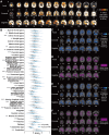
Graphical Abstract
 Figure 1
Study profile. (A) Flowchart of the timeline of the participants. (B) CONSORT diagram. (C) Infecting variants for each patient, inferred from the date of infection and genomic surveillance data from the National Institute of Infectious Diseases in Japan. (D) Number of cases with insufficient or sufficient vaccination before infection. Abbreviations: SUVR, standardized uptake value ratio; SPM, statistical parametric mapping; RBANS, repeatable battery for the assessment of neuropsychological status; jRCTs, Japan Registry of Clinical Trials.
 Figure 2
AMPAR distribution pattern in cognitive impairments in long COVID (Cog-LC). (A) Elevations in [11C]K-2 SUVRWM in patients with Cog-LC (n = 30) compared to HCs (n = 80) (P < 0.05, T > 1.66, one-tailed, FDRc). (B) Multiple comparisons across Hammers’ VOIs between HCs (n = 80) and Cog-LC (n = 30). Bold line and dashed line of each plot represents mean and quartiles, respectively. *P < 0.05, **P < 0.01, ***P < 0.001 (Bonferroni multiple comparison test after two-way ANOVA analysis). (C) Brain regions showing a negative correlation between [11C]K-2 SUVRWM and picture-naming scores of the RBANS in Cog-LC (n = 30) (P < 0.05, T > 1.71, one-tailed, FDRc). (D) Overlapping brain regions between the clusters in A and C. (E) Brain regions showing a negative correlation between [11C]K-2 SUVRWM and figure recall scores of the RBANS in Cog-LC (n = 30) (P < 0.05, T > 1.71, one-tailed, FDRc). (F) Overlapping brain regions between the clusters in A and E. Abbreviations: A, anterior; P, posterior; R, right; L, left; FDRc, false discovery rate correction.
 Figure 3
Correlation between elevated AMPAR density and plasma protein levels. (A) Brain regions showing a significant positive correlation between [11C]K-2 SUVRWM and plasma TNFSF12 concentration (n = 30, multiple regression analysis using age and sex as covariates, P < 0.05, T > 1.71, one-tailed, FDRc). (B) Correlation between average SUVRWM in a significant cluster and plasma TNFSF12 concentrations (correlation coefficient = 0.6051, *P < 0.001). Each dot represents SUVRWM of the brain region shown in Fig. 3A and plasma TNFSF12 concentration of each participant. (C) Overlapping brain regions between the clusters in Figs 2A and 4A. (D) Brain regions showing a significant negative correlation between [11C]K-2 SUVRWM and plasma CCL2 concentration in Cog-LC (n = 30) (n = 30, multiple regression analysis using age and sex as covariates, P < 0.05, T > 1.71, one-tailed, FDRc). (E) Correlation between average SUVRWM in a significant cluster and plasma CCL2 concentrations (two-tailed Pearson correlation analysis: correlation coefficient = −0.6583, *P < 0.001). Each dot represents SUVRWM of the brain region shown in D and plasma CCL2 concentration of each participant. (F) Overlapping brain regions between clusters in Figs 2A and 4D. In B and E, solid lines represent the regression, and dashed lines represent the 95% CI. Abbreviations: TNFSF12, tumour necrosis factor superfamily 12; CCL2, C-C motif chemokine ligand 2; FDRc, false discovery rate correction; A, anterior; P, posterior; R, right; L, left.
 Figure 4
Diagnostic performance of PLS algorithm. (A) Predicted values from the PLS models are displayed using a violin plot and box plot. Each grey dot represents each participant. The means of the predicted values are represented by circular points. (B) Receiver operating characteristic curve (solid line) illustrates the diagnostic performance of the PLS algorithm in distinguishing patients with Cog-LCs from HCs. The area under the curve was 0.980 (95% confidence interval [CI]: 0.960–0.999). The optimal cutoff was 0.394, providing a sensitivity of 100.0% for identifying patients (29 of 30), specificity of 91.25% (73 of 80), positive predictive value of 81.08% (30 of 37), and negative predictive value of 100.0% (73 of 73). (C) The beta coefficients from a representative partial least squares model, which are associated with disease status, are shown in the visualization. A higher beta coefficient indicates that the predicted value increases as the SUVR value of a corresponding voxel increases. As a higher predicted value suggests a stronger likelihood of Cog-LCs diagnosis, these coefficients highlight the relationship between specific voxel SUVR values and the probability of distinguishing Cog-LCs from HCs. Abbreviations: A, anterior; P, posterior; R, right; L, left; PLS, partial least squares; SUVR, standardized uptake value.
References
-
- Chen H, Zhang L, Zhang Y, et al. Prevalence and clinical features of long COVID from omicron infection in children and adults. J Infect. 2023;86(4):e97–e99. - PubMed
.png)


