.png)

Introduction
Relatively few complete medieval books have survived in the Nordic countries. In Swedish cultural heritage collections, for instance, their number runs to a little over one thousand. At the same time, Nordic libraries and archives have very large collections of manuscript fragments. This is because the parchment from medieval books once owned by the Church was systematically re-purposed by the early-modern royal administration in the 16th and 17th centuries as covers for taxation records. The number of fragments for all of the Nordic countries is ca. 50,000 from at least 15,000 manuscript books, with more than 22,000 fragments in Sweden alone, and they arguably constitute an uncommonly representative sampling of the book resources of one medieval realm1. These objects, originating mostly from books used in parish churches, represent the diversity of early book culture in the Nordic countries and Latin medieval book usage2,3. Their sheer number makes them an important body of evidence for a number of disciplines shedding light on medieval history.
Although the content of these principally standard liturgical texts is relatively well-known, at least on a general level, less is known about the history of these books as physical items. How, when, and where were they manufactured? Primarily traditional methods used in manuscript studies, such as palaeography and codicology, have been applied for the contextualization of these objects. Less has been done by way of scientific analysis. The lack of research applying methods of the natural sciences is particularly noteworthy regarding colourants, which have been very successfully studied by scientific approaches in many other countries and contexts. Our project seeks to address this gap in scholarship. It presents a pilot on the application of scientific methods to the materials—especially colourants—used in the making of manuscripts in the Nordic Countries. We have set out to examine a selected number of fragments thought to have been locally produced through technical analysis of the pigments, dyes, and other painting materials. What substances were used and how, and how does that compare to production elsewhere in Europe? How do the books compare with each other within the collection? Analyses of the palette of these locally-produced objects contribute to our understanding of their production contexts and manufacturing processes and shed light on international networks of trade and learning through which literary know-how, pigments, ink recipes, and ideas were disseminated in the Middle Ages. Our results are admittedly preliminary, due to the small number of objects studied. Nevertheless, they demonstrate the potential of such methods in the Nordic context, suggest some features that could be characteristic of the local production, and indicate avenues for further work in the field.
Background
In 2020, representatives from Nordic heritage institutions and universities met with experts in Helsinki to discuss the possibilities of investigating the materiality of the Nordic manuscript fragments using scientific methods. The meeting was part of the NOS-HS-funded project Nordic Medieval Book Culture in the European Context. Following this colloquium, a user group was formed to further the use of such methods. In 2022, an application for the project The Materiality of the Medieval North—the palette of Swedish and Finnish reconstructed Manuscripts (MoMNoPSaF) was submitted to and accepted by the European consortium Iperion HS and their mobile infrastructure MOLAB. In 2023, various analyses were carried out at the National Archives in Stockholm.
While methodologically novel, the project builds on a 100-year-old tradition of research on and cataloguing work of the Stockholm and Helsinki fragments. The fragments in Stockholm were catalogued in two lengthy projects between the 1930s and early 2000s. In the latter project, the so-called ‘Medeltida pergamentomslag’ (1995–2004, abbreviated MPO, translates ‘Medieval Parchment Covers’) an open-access database with metadata about over 22,000 fragments and images of most of them, was created. It is maintained by the National Archives in Stockholm (https://sok.riksarkivet.se/mpo). Another central outcome of the MPO project was the monograph From Manuscripts to Wrappers4. In this book, Jan Brunius summarized essential information acquired during the cataloguing process. It provides useful overviews of the dating of the fragments, the types of books they come from, texts and authors found in them, provenance and origin, and pieces together fragments that derive from the same manuscript book.
In Finland, the cataloguing of the c. 6660 fragments at the National Library started in the 1920s and is now approaching its conclusion. An important stage in this work was the project Literary culture in medieval Finland led by Professor Tuomas Heikkilä between 2008 and 2012, which also created a detailed view of the oldest literary culture in Finland5. In conjunction with this project, the Helsinki fragments were digitized and an open-access database maintained by the National Library of Finland was created. In recent years, Heikkilä has conducted pioneering experimentation in applying scientific methods such as C14 dating, parchment DNA sequencing, isotope analysis, etc. for the study of parchment6,7. The ERC-funded ‘Books of the Medieval Parish Church’ (BOMPAC) project, hosted by the National Library in Helsinki and led by Dr. Jaakko Tahkokallio, is presently continuing the study of the Helsinki and Stockholm fragments by traditional palaeographical and codicological means. From September 2024 onwards, Heikkilä’s new project ‘Combining Humanities And natural science Research to study Medieval texts, scribes, and craftsmanship’ (CHARM) approaches Finland’s oldest written culture from the perspective of literary knowhow and manuscript production. The project combines traditional humanities methods with biocodicology, and some of the imaging techniques described in this article.
Internationally, scientific methods for the non-invasive study of colourants used in medieval manuscripts have advanced significantly over the last ten years, and standardized approaches have been developed8. The scientific study of medieval manuscripts by non-invasive techniques has resulted in several publications on the materials of manuscripts, from e.g. England, France, Italy, Portugal, and Germany9,10,11,12,13,14,15,16. However, these approaches have not been applied to manuscripts in Sweden, where only colourants on polychrome sculptures, murals, and oil paintings have been investigated using scientific methods15,17.
Previous studies have shown how diverse and complex the manuscripts and the techniques employed to manufacture them are from a material perspective. Furthermore, these previous analyses have contributed to the attribution of illuminations to specific artists and provided information on the divisions of labour in scriptoria. There are several examples where scientific analysis combined with art historical and palaeographical studies have, for instance, shown the probable existence of several hands working on one volume9,10,16,18.
Methods
The aim of this study has been to identify and characterize the pigments, colourants, binding media and gilding materials, in situ, applying non-invasive methods. Expertise and instrumentation were provided through the Iperion HS platform mobile infrastructure MOLAB. The study builds upon the analytical protocol described in Panayotova 2020 with a combination of mapping/ imaging by X-ray fluorescence spectroscopy, hyperspectral imaging and point analyses by Raman and Fourier Transform Infrared spectroscopy (FTIR)8. In 2023, MOLAB experts carried out analyses at the conservation lab of the Swedish National Archives in Stockholm (Fig. 1). Prior to these analyses the selected objects had been studied by applying visual and condition examination, documented by photography, multi-band imaging (MBI), and optical microscopy studies.
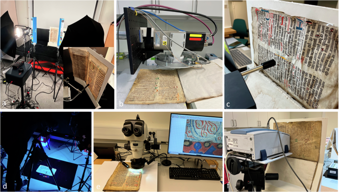
a HSI and FORS, b MA-XRF scanning, c Raman spectroscopy, d MBI, e optical microscopy, and f FTIR spectroscopy.
MBI was carried out to better visualize the colour palette to help plan and guide further analysis. Secondly, hyperspectral imaging (HSI), fibre optic reflectance spectroscopy (FORS), Fourier transform infrared (FTIR) and Raman spectroscopy, and macro and micro-x-ray fluorescence (MA-XRF and µ-XRF, respectively) scanning were conducted.
Selection of fragments and preparation
Most fragments in the Swedish National Archives are still attached as covers to the 16th and early 17th centuries taxation records. Often, the fragments are complete bifolia (two leaves). During the cataloguing process (see above), each fragment was given a single identifier—a fragment number (e.g. Fr 9635)—regardless of how many leaves the fragment consists of. Thus, a single fragment number may refer to two leaves or four pages (recto, verso). It should be noted that while most fragments remain in their early-modern account context, some have been removed from the accounts.
The ten fragments (see Table 1 and Fig. 2) were selected from those which were identified in previous research as having been certainly or very likely produced locally in Sweden. These origin attributions are mainly based on the contents, especially the presence of local saints in the liturgy, and, to a lesser extent, on palaeographical studies4. In some instances, there is also information on where the manuscript was used later in the medieval period. One fragment of unknown origin (Fr 3814) was included simply because it showed a more varied array of paints in an illuminated image. Sizes of the leaves range from ca. 27 × 20 to 37 × 24 cm. The measurements listed here document the current state of the objects with parts of the parchment folded in some cases. Thickness was measured as an average from measurements taken from three spots on the parchment. Origin, type of book, and dating were taken from the cataloguing information of the fragments (the MPO database; see introduction), except for Fr 7285, for which the origin information was retrieved from a previous study19. For three of the fragments (Fr 9635, 26692 and 7285) chosen for this project, two pages were analysed and for the rest, only one page, i.e. 13 pages in total.
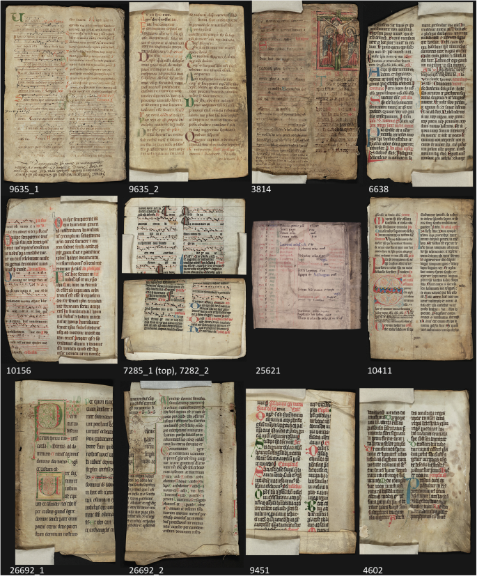
Fragment number is seen below in white. For three of the chosen fragments, two pages were analysed (Fr 9635, 7285 and 26692).
Analytical methods
Microscopy
Microscopy was carried out with a Nikon stereo microscope (Tokyo, Japan), SMZ1270i, in the range of ×6.3–×80 magnification, with an objective P-Plan Apo ×1/WF, N.A. 0.105, W.D. 70 mm, and external LED hi-power spotlighting at 5500 K. Images were captured by camera BL5.0FX264 with a 2/3” Sony Pregius CMOS-sensor with 5Mp resolution (2448 × 2048), pixel size 3.45 × 3.45 μm. The acquisition software was Nikon NIS-Elements 5.20.02. No filters were used.
Multiband imaging (MBI)
All images were acquired using a modified full spectrum Nikon D850 camera (internal IR–UV-blocking filter removed) with a Nikon AF Nikkor 50 mm lens. The objects were placed flat approximately 100 cm from the camera. Two identical light sources were placed symmetrically at approximately 45° angle toward the objects. Images in the visible range were acquired with two Godox V860ii speedlights and a CHSOS Robertina UV–IR blocking bandpass filter. Infrared reflection images were acquired with two Godox V860ii speedlights and a B + W 093 IR cut-on filter. UV images were acquired using two CHSOS Fabrizio UV lights and CHSOS Robertina UV-IR blocking bandpass filter (UV luminescence) or a CHSOS VIS-IR blocking filter (UV reflectance). A Calibrite colorchecker and Spectralon diffuse reflectance standards (2%, 50%, and 99%) were used for calibration. A white, non-luminescent homogeneous paper board was used for flat-fielding. Postprocessing was completed following the guidelines proposed in the user manual and nip2 software20. Full data set and technical specifications are available on Zenodo (https://doi.org/10.5281/zenodo.10869945).
Hyperspectral Imaging in visible and UV fluorescence (385–1040 nm)
For hyperspectral imaging, a SOC710 produced by Surface Optics Corporation (San Diego, USA), was used. The system utilizes a whiskbroom line scanner producing a 696 × 520 pixels hypercube covering the range of 400–1000 nm with 128 spectral bands. The spectra are defined by one point approximately every 4.5 nm. The spatial resolution can be continuously modulated by the adjustable focal length of the mounted objective, reaching about 300 µm in this set-up. Two Elinchrom Scanlite 350 W Halogen lamps with diffusing umbrellas were used for reflectance measurements while two Honle Ledline 500 led systems emitting at 405 nm were used for luminescence measurements.
Macro X-ray fluorescence imaging (MA-XRF)
The Macro X-ray fluorescence (MA-XRF) scanning technique was carried out using the mobile XRF scanner developed at the XRAYLab laboratory of the Istituto di Scienze del Patrimonio Culturale—Consiglio Nazionale delle Ricerche (ISPC-CNR) of Catania21. The mechatronic scanning system, developed in real-time technology, consists of a spectrometric measuring head based on a microfocus X-ray source with a 30 W Mo anode coupled with a focusing polycapillary lens (minimum beam size of 50 μm at 1.5 cm from the analysed surface). X-ray fluorescence, induced by primary radiation, is detected in event mode by an ultra-compact multi-detector system consisting of 4-SDDs (silicon drift detectors) operating in parallel, each with an active area of 50 mm² collimated to 40 mm² resulting in a total active area of 160 mm² with an energy resolution <130 eV at 5.9 keV. The 4-SDDs are arranged around the polycapillary lens on a circular array mounted on the primary source, achieving a 90°–45° (incident/detection) geometry with respect to the sample surface. The spectrometric head is mounted on a 3-axis system (XYZ) with a stroke of 50 × 50 × 20 cm³, enabling continuous scanning at high speed (up to 50 mm/s). The scanning system is fully controlled by a control processing unit (CPU) developed in a multi-node design and programmed in a real-time environment. Users enter and monitor all measurement parameters during scanning through an interactive dashboard. The CPU performs dynamic analysis of all pixel-spectra using an accurate fit procedure, providing the elemental images in real-time without artifacts during scanning. Additionally, the scanning system is equipped with in-house programmed software for real-time image analysis, allowing correlation of images (RGB), application of logical-mathematical tools, statistical analysis (e.g., PCA, ICA, and NNMF), generation of scatterplots, and export of individual XRF spectra by selecting any area (ROI) of the image. The pages under study have been fully mapped, and the full area of each fragment was covered in less than 2 h (see Table S1) at 20–25 mm/s scanning speed, corresponding to a pixel size of 400–500 µm and a dwell time/pixel of 20 ms. Additionally, some decorative details, inks, or capital letters were analysed at a higher lateral resolution of 100 µm (µ-XRF mapping) and a dwell time/pixel of 20 ms (scanning speed 50 mm/s). See supplementary information, SI1, for a comprehensive list of the fragments analysed by MA-XRF and µ-XRF, with an indication of a number of steps to analyse the whole area, step size, and total acquisition time. Due to the fragility and physical size of the volumes, the MA-XRF scanning was carried out by assembling the scanning system to operate in a horizontal position. To ensure that the elemental mapping of a single page did not include fluorescence signals from the underlying pages, a dedicated support was constructed. The support (45 × 30 × 30 cm) was made of 2 cm-thick expanded polyethylene, with a central rectangular hollow area where a thin polyester sheet (0.1 mm thickness) was placed to hold the page being mapped. While the presence of the polyester sheet introduced higher coherent and incoherent scattering signals, it enabled the mapping of individual pages without contributions from the underlying ones.
Fourier Transform Infrared spectroscopy (FTIR) mid-region
Investigations were carried out utilizing a portable Bruker Optics (Billerica, Massachusetts, USA) ALPHA-R spectrometer equipped with a Globar infrared radiation source, a Michelson interferometer (RockSolid(TM)), and a DLaTGS detector. Positioned at a distance of 1 cm from the surface under examination, data was collected in external reflection mode with an angle of incidence of light at approximately 20° where the investigated sample area width, as determined by the probe diameter, was about 4 mm. The video camera permitted the point of analysis to be visualized and monitored and spectra were gathered in the near/mid-FTIR range from 7500 to 350 cm−1, observing a spectral resolution of 4 cm−1 with a spatial resolution better than 7 mm2.
Raman spectroscopy
The portable Raman i-Raman Plus by B&W TEK (Plainsboro, NJ, USA) equipped with two laser sources at 532 and 785 nm and two dedicated CCD detectors was utilized in this work. Measurements were captured with an optical fibre probe on 90–100 µm spot. Spectra were acquired across the range 65–4200 cm−1. Laser power was modulated by software (2–6 mW for the 532 nm and max. 12 mW for the 785 nm excitation) in accordance with the area under investigation.
Results and discussion
The selection of fragments contained many of those pigments already noted in the extensive analytical work conducted elsewhere in Europe7,8,9,10,11,12,13,14, such as azurite, red lead, vermilion, orpiment, and copper-based greens. Even though these fragment pages might appear rather simple in terms of their colour scheme, the analysis has shown a greater variation and complexity than first meets the eye. This observation will be further elaborated below where findings are listed by colour. A summary of the findings can be found in Table 2. Further images and XRF maps can be found in Supplementary information 2 (SI2), and 3 (SI3).
Blue
All blues, except one, were identified as azurite by HSI (absorption max 640 nm) and FTIR (a strong doublet at 4380 and 4244 cm−1, which can be attributed to both the combination of ν + δ (OH) and the overtone 3ν3. This doublet partially overlaps with the methylenic C–H stretching and bending combination from lipidic binders), and is further corroborated by the presence of copper in the XRF analysis12,22. Azurite is a basic copper (II)-carbonate: 2 CuCO3·Cu(OH)2 forming bright blue crystals. The pigment can be prepared either from naturally occurring minerals or produced synthetically. The presence of particular impurities can arguably be used to attribute origin and identify leaves that come from the same workshop23. The azurites of the pages of the investigated selection show a variation in impurities, which could indicate different geological sources or different grades of purification. They showed the presence of barium, iron, zinc, arsenic, manganese and bismuth in XRF analysis with notable variability between the various fragments (Fig. 3, Table 2). The combination of impurities can thus confirm the likeness or not of the different fragments or various leaves of the same fragment. The presence of impurities in azurite has been noted in other analyses. For example, iron has been observed in 11th-century French manuscripts and zinc, arsenic, and barium in one 16th-century Flemish Book of Hours24. Barium, arsenic, zirconium, and bismuth were detected in two Italian manuscripts from the 13th and 15th centuries while antimony and silver were found in a 15th-century Spanish manuscript in synchrotron XRF analysis23.

Micrographs (a) of azurite blues of four fragments and XRF spectra of the blue areas shown in SI2 (b). Note variability in minor components such as manganese, barium, zinc, arsenic, and bismuth.
Azurites are described as rendering varied colour hues depending on grain size16. The azurite blues of the selection show a variation of finely ground particles hardly discernible at ×80 magnification (Fr 4602 and Fr 10156) to a coarser grain size of ~20–40 µm in Fr 6638 and Fr 3814.
The exception to the use of azurite was the blue writing in the 12th-century calendar fragment Fr 25621, which was identified as ultramarine with HSI, (absorption max 600 nm)12, and Raman spectroscopy (peak located at 545 cm−1 attributed to ʋsym(S3−)25 (Fig. 4). Raman band at 1316 cm−1 indicates the possible presence of natural mineral such as diopside (CaMgSi2O6), commonly associated with lapis lazuli in nature26, and suggests that transition metal dopants in the diopside may be responsible for the Raman features, likely the result of fluorescence with vibronic coupling27. Ultramarine is not an uncommon pigment used for manuscript colours. The source of natural ultramarine is the rock lapis lazuli, which contains a mixture of the minerals calcite, pyrite, and lazurite. The latter is a complex sulfur-containing sodium aluminium silicate, Na6Ca2(Al6Si6O24)S2, which is the actual colouring agent of ultramarine28. This pigment has been found in many manuscript illuminations but was not so commonly used as ink to write text. However, blue text is quite often encountered in calendars. While the colour red was the most common one used to designate the most important saints29, blue was also used, at least in twelfth- and thirteenth-century manuscripts.

Fragment 25621 (a, detail), a 12th-century calendar with blue text identified as ultramarine. b Microscopic view (×80 magnification), c Raman spectrum (exc.785) and d HSI mean spectrum (blue line, absorption max. 600 nm) with standard deviation (blue area) and ultramarine reference spectra (pure, in powder, dotted black line).
Natural ultramarine is described as a precious pigment. The preciousness of ultramarine stems from the scarcity of known mineral sources for the lapis lazuli rock from which the pigment is obtained. Probable places of origin are Tajikistan, Pakistan, and Afghanistan26. It has been observed that there was a varying geographical and chronological use of different blues in the early medieval period, as azurite is more commonly found in Carolingian manuscripts, and ultramarine appears more often in Ottonian manuscripts and in early medieval England. In England, the use of azurite was not common until the 12th century9,10. The calendar fragment, Fr 25621, was produced in the late 12th century. The only Nordic saints in the original hand are Olav and Canute, which points to the calendar originating in the diocese of Linköping or possibly Denmark. Certainly, the manuscript was later adapted for use in the diocese of Linköping. As for ultramarine’s other applications in the Swedish context, the use of this pigment in contemporary Swedish wall paintings was not very common; it was only found in 10 out of the 70 analysed medieval church murals30. The blue text of Fr 25621 is also associated with the presence of lead, with lead white being confirmed by external reflectance FTIR (SI4_1), for the characteristic combination bands (CO3−2) at ca.1730 and 2400 cm−1 22.
Red
Not surprisingly, red is the most common colourant as it is used in rubrication. Out of 17,740 colour entries in the MPO database, 36% are listed as containing red, compared with 29% green, and 24% blue, while other colourants are below 10%. Most reds in the selection are identified as vermilion—the mineral cinnabar, HgS. Cinnabar was confirmed by Raman (main band 255 and 341 cm−1) for 28 out of 36 listed red and verified in HSI (inflection point at 600 nm) as well as indicated by the presence of mercury (XRF). The mineral cinnabar is usually associated with mercury mining in the Mediterranean, Central Asia, and the Pacific Ocean. Only a few places in the world have been historically relevant for its exploitation as a pigment, the known European ones being: Almadén (Spain), Idrija (Slovenia), Mount Amiata (Italy), Génépy (France), and Moschellandsberg (Germany)26. Hence, the presence of cinnabar in the analysed fragments constitutes evidence of material import. The term “vermilion” is used for the synthetic version of HgS, which, considering the period, is likely to be the one used in these fragments, and henceforth used for this red pigment. For some of these reds in initials and rubrication (in Fr 4602, 6638, 7285, 10411, 26692), lead was also detected by XRF indicating a mixture with a lead-based pigment. This was deduced to be red lead from the observation of characteristic peaks in Raman spectra (122, 391 and main 550 cm−1 due to stretching of the Pb(IV)–O-bond) of this pigment) and the lack of characteristic peaks for lead white in FTIR spectra, thus excluding the other common lead-containing pigment31. Mixtures of red lead and vermilion have been found in previous analyses of pigments in manuscripts10,14,16. Indeed, they are the most commonly encountered reds in the rubrication of medieval manuscripts. In fragments Fr 9451, parts of Fr 10411, Fr 10156, Fr 25621, and Fr 3814 (further discussed below) vermilion is the only red pigment present. In Fr 9451, the detection of sulfur in red areas is also associated with sulfate, as gypsum was indicated by HSI and FTIR (SI4_2). Calcite (sharp FTIR combination bands around 1800 cm−1; 2510 and shoulder 2594 cm−1), was also found in some reds used in Fr 10411 and Fr 26692 together with vermilion and it seems to have been used to adjust the hue (SI4_3 and 4).
The exceptions to the mercury-based reds are the several reds of Fr 9635, where upon visual inspection, three to four different reds can be seen (Fig. 5, SI2_1 and 2). There is light red for the rubrication of ‘e’, ‘a’, and ‘F’ in the upper left corner and, the red text; the dark red initials ‘Q’, ‘D’, and smaller ‘C’, and then the medium red of large ‘C’ (top right). To map the distribution of the different reds, Spectral Angle Mapper (SAM) analysis has been performed taking into account the HSI spectra (in the range 450–970 nm) of the reds used for the capital letters (Fig. 5). The mapping shows that the light red used for the ‘F’, on the top left (in yellow in Fig. 5) was also used for part of the text. The dark red used for the ‘D’, in the left (in red in Fig. 5) was also used for the small ‘C’ and the ‘Q’ in the next column. Finally, the red used for the larger ‘C’ on the right (in magenta in Fig. 5) presents significant similarities with the light red spectra. The light red showed only lead in XRF analysis (SI2_1), suggesting the presence of red lead, also corroborated by Raman spectroscopy (characteristic bands at 120 and 550 cm−1) and by the HSI spectrum with an inflection point at 570/560 nm (Fig. 5) (SI4_8). Raman analysis also shows the presence of massicot in red text (with the presence of ulterior bands in addition to red lead at 140 and 288 cm−1)10,31,32.

Plot of HSI spectra of the reds in the 450–970 nm range (b) and their distribution in the manuscript (a). a Light red pigment (yellow curve in b), medium red (in magenta), and dark red (in red) (b). Micrographs of light red ‘F’ (c), dark red ‘D’ (d) and medium red ‘C’ (e).
Both lead and iron are present in the dark reds, and Raman analysis indicates a mixture with red lead. The presence of iron is stronger for the darker red ‘Q’ and ‘D’. The absorption maximum at 850 nm in the FORS spectrum in Fig. 5 is indicative of a red ochre7. Interestingly several other elements can be found in particles in the red ink of these letters, namely zinc, copper, and manganese (SI2_2). Particles containing mercury are also detected in the large ‘D’ (SI2_2). In the medium red capital ‘C’ XRF analysis detected particles containing zinc, copper and manganese similar to those seen in the dark red areas.
The presence of impurities indicates a not very finely processed pigment. Fr 9635, which dates from the second half of the 12th century, is one of the earliest fragments identified as being of Swedish origin. It is thought to have been used in the diocese of Uppsala. A visually similar red, also containing copper particles, has been observed in the XRF analysis of a later Swedish law text, Östgötalagen MS B 50, dated to ~1325–1375 (personal communication Evans and Norrehed). Copper in the same mineral locality as iron is, for instance found in the Falun copper mine in central Sweden, where small-scale copper mining probably was practiced as early as in the 10th century33. Iron oxide, a by-product of copper mining, is known to have been used to produce a red paint, also containing copper vitriol. The pigment is made from heated haematite. In addition, Sweden has one of the earliest examples of the production of iron from iron ore, situated in Norberg in Bergslagen33.
Another fragment, Fr 3814, showed a varied use of the reds in the Christ image: two hues of red appear in the drapery of the clothes, in Christ’s halo, and in the frame. Furthermore, there is the red of the wounds and a pinkish modelling of the body of Christ (SI2_3). From XRF scans, it is clear that vermilion has been used for the dark red shading of the drapery and the darker side of the frame, while the lighter red parts are vermilion mixed with red lead. The red for the rubrics and initials is also vermilion and red lead, a combination similar to that seen as common from the 12th century onward in British manuscripts9. In addition, XRF, FTIR and Raman analyses show that the hue of the light red of the clothes and drapery has been made lighter with calcium carbonate, which is also the main component for the flesh tones of Christ’s body. Most common flesh tones in medieval illuminations are lead white, often in combination with vermilion9, while here calcium carbonate is used as already documented in several examples9,10. For the wounds, only vermillion has been used.
The pinkish brushstrokes modelling Christ’s body did not reveal any identifiable elements in XRF analysis, possibly indicating the use of an organic colourant as these were commonly employed in manuscripts for pink glazes10,34. This hypothesis of an organic colourant can neither be confirmed nor rejected here because while the shape of the reflectance spectrum did not have those of an anthraquinonic dye, a different organic red material cannot be ruled out (SI4_9).
Green
There were various colourants and pigments as well as their mixtures to render green colours in medieval manuscript production. Various copper compounds have been used, such as verdigris (a collective name for several copper acetates), copper sulfates and malachite, a basic copper carbonate. Often a mixture of a blue and a yellow has been used, for example, azurite and orpiment, or indigo and orpiment10. There are also entirely organic green colourants, such as sap green, that have been used10. More greyish hues are possibly given by green earth, a mixture of aluminosilicates of iron, magnesium, aluminium, and potassium (the colouring agent being a mineral such as celadonite or glauconite), but other minerals can be present. Green earth has been particularly connected to Byzantine and northern Italian manuscripts10.
Green appears in six of the fragments examined in this study, showing a variation in hues ranging from dark to very light. All but one show copper in XRF analysis. In the analytical non-invasive investigation performed, it has not been possible to differentiate between the various copper-containing varieties. None of the greens shows clear spectral characteristics of malachite in FTIR, although bands of carbonate are observed. It is possible, however, to see the characteristic strikethrough on the other side of the parchment page normally associated with copper pigments for Fr 10156, the bright green of Fr 9635, the various greens of Fr 26692, 25621, and to a lesser extent, for Fr 4602. For 10156 and 26692 verdigris could be suggested by carboxylic stretching peaks possibly relative to basic copper acetate at 1556 and 1450 cm−1 (Fig. 6). For Fr 3814 it is not obvious, and there is no strikethrough for the dark green of Fr 9451 although FTIR also shows the presence of silicates in the 1000 cm−1 region (beyond parchment signals), not excluding the hypothesis of chrysocolla, a hydrous copper phyllosilicate35. The FTIR results for Fr 9451 also show a particularly strong presence of calcium oxalate (highlighted bands ca. 780 and 1610 cm−1)36 and an organic component of lipidic origin not excluding a wax given the characteristic inverted CH bands in the region 2930–2850 cm−1 37.

Fr 9451 (a), micrograph (b) and FTIR spectra of the greens of Fr 10156, 26692 and 9451 (c). Dotted line indicate peaks for the presence of silicates for 9451 and 26692, and for carbonate for 10156 and 26692, asterisk show bands for calcium oxalate and arrows indicate carboxylic stretching peaks possibly related to basic copper acetate.
Apart from copper being detected, other elements associated with the green are zinc in Fr 26692 and chlorine in the green of Fr 10156. Chlorine has been associated with a peculiar element of the salt used in the production process of the so-called ‘salt verdigris’, where copper is enclosed for a number of days coated with honey, salt, and vinegar38. It has been identified in the green colour of, for instance, a German illumination from the 16th century, and in some greens in the 12th century Bury Bible, from Bury St. Edmunds, England10. Verdigris, high in zinc, has been found in a gospel book from England possibly from the 11th century10.
The only green not containing copper is the dull greyish green in the initials of Fr 9635 (small and big ‘A’, ‘D’, ‘p’ and ‘C’, SI2_4). The grey-green shows a clear presence of iron in combination with manganese, potassium, calcium and phosphorus (see SI2_5). FTIR suggests the presence of calcite, silicate, where in only one point, in small A, signals also of haematite can be observed (bands at 540 and 467 cm−1). The presence of iron, potassium and silicate could suggest the presence of green earth, but the diagnostic features of caledonite and glauconite were not observed by FTIR and no evidence for the presence of this pigment is seen in the vis-hyperspectral; at least not on the surface of the green shade. Whilst the presence of phosphorus could indicate a mix with bone black, the characteristic presence of bone black was not, however, observed by FTIR.
Yellow
Yellow is a less frequent colour on the leaves of these more text-dominated manuscripts. For this selection, yellow is found in five of the fragments. In our analysis, yellowish transparent layers, encountered in two manuscripts (Fr 26692 and Fr 9635, Fig. 7), have also been grouped together with more clearly yellow components. Usual yellows in medieval manuscript paintings are composed of arsenic sulfides such as orpiment, lead-tin yellow, or iron hydroxides (ochres). Organic colourants such as weld, safflower, saffron, and buckthorn were also used9,10. Evidence of these organic colourants was not observed with the non-invasive techniques applied in the present study. However, it was possible to note the strong co-presence of calcite in these areas, which may support the hypothesis of an organic yellow which could well have been absorbed or precipitated on calcite for the preparation of the pigment lake (Fig. 7a bands of calcite highlighted therein)39. Almost transparent layers are visible in Fr 26692 and 9635, in which no element but calcium can be determined by XRF, and no discerning identification could be made by the techniques utilized here.
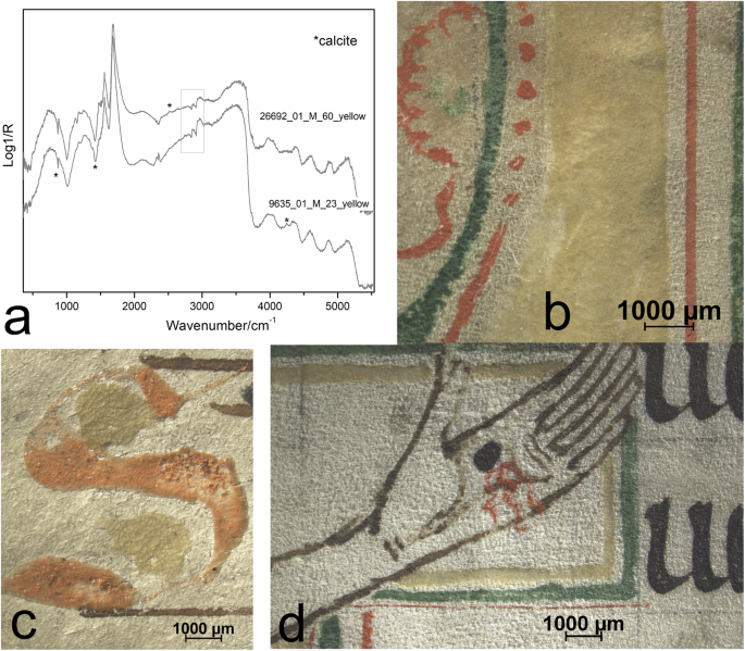
FTIR spectra (a) and micrographs of parts with yellowish transparent layers in Fr 26692 (b, d) and Fr 9635 (c). It is possible to note the co-presence of calcite in these areas which may support the hypothesis of an organic yellow.
The yellows found in this selection range from bright yellow to almost all transparent. Fr 6638 shows an unusually bright and opaque yellow. This yellow was identified as orpiment because of the high scattering of arsenic sulfide compounds in the Raman analysis (main bands for orpiment- 152, 314, 355 cm−1) (Fig. 8) and arsenic clearly showing in the XRF scans (SI2_7)32,40. It is also possible to see particles of a more orange hue possibly indicating realgar (additional band Raman 270 cm−1) (Fig. 8). It is not uncommon for these arsenic sulfides to occur together. Orpiment was likewise identified in the yellow of the halos and Mary’s arm in the crucifixion scene of Fr 3814, and in the yellow flourishes of Fr 10411, all showing a more subdued colour than the yellow of Fr 6638. In the halos they are probably mixed with calcium carbonate, which was identified by FTIR and HSI, corroborated by a clear detection of calcium in XRF for this pictorial part (SI2_3).

Yellow of Fr 6638 (a, b), identified as orpiment. Orange particles of possible realgar/pararealgar are seen in micrograph (b) and by Raman spectroscopy (c).
Gold
Gold can be seen in two of the objects, Fr 10411 and 3814, and its presence is verified with XRF (SI2_6 and 8). In Fr 3814, it is in the background of the image, the whole of it being most likely covered with gold on an earth-pigmented ground (SI2_8), as iron was also detected by XRF for this field. In Fr 10411 gold is in the dots of the decorated initial ‘S’ (SI2_6).
In Fr 10411, the gold has a yellow base layer of orpiment. The use of orpiment together with gold has been noted in other studies9,14. In 12th-century English manuscripts, orpiment was used on top of gold9. Although it is tempting to speculate that the gold was added at a later stage due to the lack of precision, this conclusion is contradicted by the visible intermixing of the gold with orpiment.
Black and brown, inks and flourishes
For all but one of the included fragments, the main bodies of text showed a significant amount of iron in the XRF analysis, allowing the identification of iron gall ink, the exception being the brown ink of Fr 9635 (SI2_9). Furthermore, the infrared reflectance of the script of Fr 9635 was transparent, indicating it is not a carbon-based ink either. The brown colour could suggest a sepia ink; however, it has not been possible to see any indication of this in FTIR. The natural habitat of the source for making sepia ink is not found in close proximity to Scandinavia; however, imported ink is feasible. Nevertheless, it is more likely that it is a tannin ink without iron, i.e., a solution of plant extract from tree bark or gall nuts are known to have been brown in colour and being transparent in infrared reflectography from 750 nm41,42,43. This seems to indicate ink preparation taking place in circumstances in which iron sulfate (green vitriol) was not present.
According to previous studies, carbon-based ink was often the preferred ink for drawing contours9,10, which is the function it has been used for in Fr 3814, shown in IR reflectance in SI2_11. It is also shown in the black flourishes of 10156 in IR reflectance (SI2_10). In Fr 26692, however, the outline of the figure of Jesus showed both iron and lead. Lead is not associated with iron, which can indicate the use of a lead stylus in sketching before applying the ink (SI2_11).
The analyses make it possible to differentiate between different iron gall inks used in the same document. This can be seen in the IR reflectance image (SI2_11, b2), where the text next to the figure of Jesus in Fr 26692 is visible in IR reflectance while the other text is more transparent. In both texts, iron is detected in XRF analysis (SI2_11, b3). The visibility of the ink next to Jesus in infrared reflectance could suggest a mix of carbon-based ink and iron gall ink.
Other instances of inks of different compositions in the same document can be seen in 10411, 6638 and 9635. In Fr 10411, an addition contains more copper than the main text, as shown by the false-colour XRF map where red is assigned to zinc and green to copper (Fig. 9e). Fr 6638 has an interlinear addition in different ink, and in Fr 9635 a different ink is seen for the musical notation. In the calendar of Fr 25261, there are visibly different black and brown inks, but it has been difficult to find any major differences between them in the analysis results. Only two brownish segments of iron are not visible in the XRF image (the writing below the red ‘Bartholomei’ and ‘augustini’ lower right, Fig. 4). It is possible that a greater resolution and a more precise Raman or more detailed infrared reflectance analysis up to above 1400 nm would be needed in order to differentiate these inks41,44.

Visible-light image of Fr 10411 (a) and µ-XRF maps of ink with interlinear addition. The iron gall ink (map of iron, d) of the interlinear added text contains copper (b) while the original text contains zinc (c). e Shows a false colour image where red is zinc and green is copper.
The analysis cannot only be used to verify different inks within the same document; it can also be applied to detect similarities in the composition of the used substances in different fragments. For instance, a very similar combination of elements other than iron can be detected by XRF in the iron gall inks used in Fr 10411 and 10156, i.e. zinc, potassium, calcium, and manganese. In order to group inks based on composition the ratio of impurities to iron could be used43.
Although medieval Swedish written sources containing recipes are scarce, recipes for ink have been found in five manuscripts with a late-medieval provenance in the Bridgettine monastery of Vadstena45. In one manuscript, probably originally produced in Germany in about the 1430s, the production of iron gall ink is described. In this recipe, gall-nuts are placed in water with acetic acid in a copper pot, after which vitriol and gum arabic are then added46. This recipe is similar to others in common use at the time. The inks used for the headings of the taxation accounts from the 16th and 17th centuries often contain zinc, an element also detected in other iron gall inks of Scandinavian origin from the same period47.
Binding media
Interference from the parchment can make an interpretation challenging in FTIR, as protein bands of parchment can overlap with signals from suspected protein-based binders. Egg-based binders, namely, egg yolk, egg white, and whole egg produce complex spectra in external reflectance FTIR with variations influenced by the paint layer’s chemical and physical properties. The key chemical difference is that egg white is purely protein-based, while egg yolk and whole egg contain a lipid fraction. As a result, their spectra not only show the amide bands but also distinct lipid features. The presence of lipids is generally identified by a sharp C = O asymmetric stretching band, typically appearing between 1800 and 1700 cm−1 with a derivative-like shape, centred around 1740 cm−1, which in this case is covered by the amide signals of the parchment. It is at this point that instead in the NIR region that the combination bands ν + δ(CH), centred at 4399 and 4266 cm−1, are discernible in both pure egg yolk and whole egg binder systems11. Given such, it is difficult to diagnose proteins from egg white, which was a commonly used binding medium. Nevertheless, the fact that lipids could also simultaneously be detected in the NIR region in many colours could even indicate the use of whole egg as well as egg white as given, for instance, for the red of initial ‘S’ in Fr 10411 (SI4_3). In several fragments, oxalates could be seen by FTIR, which could be an indication of binding medium degradation.
Observations on technique and divisions of labour
Inks and initials
In a scriptorium, there was often a division of labour among different specialized craftsmen with regards to copying the text, illuminating, and adding musical notation48. It has been suggested that this specialization was not very developed in Sweden49. In this study, it has become evident that inks of the same colour but with varied functions on the page often differ in composition. In our interpretation, the variation in the inks suggests a division of labour. While the possibility that the same craftsperson used different raw materials for different inks during various stages of book production of the page, such as copying the text and decorating, cannot be completely ruled out, this appears the most simple reading of the evidence. Some examples of these variations are described below.
For Fr 10411, the blues of the minor initials in the text and the blue of the major initial ‘S’ show a different composition in regard of barium content in relation to copper, barium being more abundant in the ‘I’ (Fig.10), possibly suggesting the use of different azurite batches. Although impurities like arsenic and zinc are present in all the letters painted in azurite, the scatter plot (d) in Fig. 10 gives evidence of two different correlations between barium and copper for the two blue initials. This result possibly implies the use of two different azurite batches. Analogously, in Smieska et al. the study of the ratio of barium to copper showed the use of at least four types of azurite on the same manuscript page and the authors suggested that this is the result of a use of different natural sources of azurite, or of a purification process23.

The flourishes and initials ‘I’ and ‘Q’ is in green square and the blue of the decorated initial ‘S’ is in yellow square. Comparison of the VIS-light image (a) and MA-XRF results: maps of barium (b) and copper (c), false-colour scatter image (e) from the scatter plot in (d) representing areas where these elements correlate, and Ba content is lower (yellow) or higher (green).
Fr 10411 also has reds with different compositions. Vermillion with red lead was used for the red of the initials and for rubrication, while for the decorated ‘S’ initial only vermilion was used, possibly in a mixture with calcium carbonate, as indicated by FTIR (SI4_3), and calcium is evident in XRF mapping.
Musical notation
There is a musical notation in three of the selected fragments. They are a gradual responsorial for Christmas (Fr 7285), a processional antiphon for Easter (Fr 9635) and an invitatory for Palm Sunday (Fr 10156). In addition, there are stave lines without notation in Fr 3814. For Fr 10156 and Fr 7285, the results do not show any evident differences between substances used for the notation and text elements. As mentioned above and visible to the eye, the colour of the notation and text of Fr 9635 is different.
All of the stave lines, apart from that of Fr 9635 are red in colour, with results showing vermilion and red lead for all reds apart from that of Fr 10156, in which only vermilion is present. The stave lines of Fr 9635 are brown but do not provide the same results as with the brown text, since lead is detected in the XRF imaging (SI2_12). This could indicate the use of brown earth or could possibly be traces of some sketching with a lead stylus. In addition, in Fr 9635, the notes of the music show a different reflectance in IR than the stave lines and the text.
Ruling
For all but one page of the two analysed pages of Fr 9635, ruling is visible on visual inspection to a greater or lesser extent, and most clearly so in Fr 9451 and Fr 7285. For three of the fragments (Fr 7285, Fr 3814 and Fr 10411) iron gall ink is indicated as the lines, either all or partly, show iron in XRF imaging. A use of lead is indicated for Fr 26692 which is consistent with the possible use of lead stylus for the contour of Christ. Interestingly, one side of Fr 9635 shows the ruling while the other side does not. The page with the ruling has been ruled with a metal instrument and lead is seen in the µ-XRF map (SI2_5). On one side, the trace of this instrument is visible, and on the other only, the indentation. The use of a metal tool for ruling was standard practice in the 12th century. For the other fragments, no conclusive results have been obtained. It can, however, be noted that the ruling seen in Fr 10156 is semi-transparent upon visual inspection, and possibly made by indentation.
Discussion and future research
Most of the colourants detected in the analyses, with some exceptions, such as Fr 9635, are common pigments in more southern European manuscript illuminations, and this study’s results suggest the import of colourants from more southern and western Europe. Written book culture arrived in Sweden together with Christianity. The most important regions from which specialists in religion and writing are known to have arrived are Germany, Denmark, and England. More generally, the best-documented trade relations of Sweden were with the Hanseatic cities and, to some extent, Northern Russia30. By the 13th century, Swedish priests were also travelling to study at universities in, for example, Paris and Bologna45. It is likely that skills and materials used for making books were transported via all of these routes. Interestingly, pigments such as ultramarine and orpiment have been described as uncommon pigments for Scandinavian church murals30, but it would seem that they were more commonly used in book production, which was obviously a different craft. Different pigments are more suitable for one medium than for another, which may be one explanation for the observed. Furthermore, the quantities of pigments needed for book decoration would have been smaller than those needed for, e.g., wall paintings.
In comparison to more southern European manuscripts, other differences may also be noted. Dyes that in several studies have been identified in illumination, such as indigo in blue or green or madder for red were not found in the analyses. The only possible organic components encountered in the study were the yellow/transparent pigments of Fr 26692 and 9635, the brown/purplish glaze of the blue azurite, and the pinkish modelling on Christ in Fr 3814. These can be of interest to investigate further with other techniques. Whether this absence of quite common substances reflects a reduced need for a diverse palette or a less developed production and trade of certain colourants is impossible to say presently. The fact that many of the analysed materials in the studied selection are in initials and rubrication, where the need for modelling and drawing is less than for illuminations, could also contribute to these results. In previous analyses of church murals, a relative lack of indigo has also been noted50. The sample selection of fragments from ten books limits the level of generalization possible. Therefore, in order to see general trends more fragments would need to be studied.
An object worth investigating further would be Fr 9635, with its several greens and reds, and an unusual ink. Saints mentioned in the text are Olav, Botvid, and Eskil the two latter of which are associated with the Mälar region of Sweden and especially the province of Södermanland. In the fragment catalogue of the Swedish National Archives, the fragment has been catalogued in MPO as coming from the same manuscript as Fr 8656. In a recent article, it has been demonstrated that also a third fragment, F.m. I.27, in Helsinki, National Library of Finland, comes from the same book51. As previously mentioned, all these fragments have been preserved by being recycled as covers for tax accounts in the 16th or 17th century. Fr 9635 became associated with Finnish accounts of 1556–1558, and F.m. I.27 was also attached to Finnish accounts (it is no longer known which one). At the same time, Fr 8656 was attached to accounts from Kalmar (Småland), situated on the southeast coast of Sweden. It would be of further interest to in the future also carry out a technical analysis of these two fragments, Fr 8656 and F.m. I27. This pattern of recycling—association of leaves from one manuscript with accounts from different parts of the realm—often indicates that a manuscript was being recycled as covers in the Central Chamber in Stockholm. Books used in the Chamber often came from the surrounding regions, i.e., Södermanland or Uppland. Given the presence of Södermanland saints and the probable later Stockholm provenance, it seems likely that the book originated and was used in the Mälar region- probably Södermanland—in the Middle Ages. One of the most promising avenues to pursue would be an examination of material of similar date (12th century) with the same kind of later archival provenance, i.e., fragments associated, in the 16th century, with records from Södermanland or those produced by the Stockholm central chamber. It would be of interest to see if other books written in tannin ink without an iron component could be identified. This could indicate book production taking place without access to green vitriol, in the same or similar context as Fr 9635. In order to screen many fragments, the protocol for ink categorization suggested by Rabin can be adopted with NIR imaging and XRF43.
Further study of the other colourants could also be helpful in trying to connect manuscripts with particular writing centres. Applying the non-invasive methods used in the present article, it could be possible to see whether similar materials have been used in two manuscripts. The presence of particular impurities in colourants, which appear in several manuscripts, could be a particularly useful indicator. Further work on the topic would, moreover, benefit from adding micro-sampling to the methodological repertoire. This would make it possible to determine organic constituents. It is possible that organic colourant materials were less often imported, and it might thus be more likely to find local substances. Pinpointing a geographical origin of the materials through these non-invasive methods has at present not been possible; therefore, and for this purpose, a micro-sampling might lead to more in-depth results. In order to determine locality, other aspects of manuscripts could be more suitable, such as determining the microbiome of the parchment, or other biocodicological study methods.
Conclusions
For the first time the palette and materials of thirteen medieval parchment leaves (from ten fragments) thought locally produced in Sweden from the 12th to the 15th century have been studied by non-invasive methods for chemical and physical characterization. The studied fragments originate mostly from books used in parish churches of the medieval Swedish realm (including present-day Finland). They could, in other words, be said to represent the everyday book culture of the Middle Ages. The results of this study reveal the presence of many of the materials commonly used in European book production at the time, such as vermilion, red lead, azurite, orpiment, and copper-based greens, and also some rarer substances.
Two fragments dated to the 12th century show different materials in comparison to others in the selection of fragments. They contain, for example, ultramarine used for the text in a calendar, a varied use of different reds and greens, and a tannin ink manufactured without iron-containing substances. For the greens, there is both copper-based green and iron-based green, tentatively identified as green earth. For red initials, the presence of iron associated with particles of zinc, copper, and manganese can be seen.
The colourant components of the studied selection evince significant variation, and it appears likely that they were received via trade routes and learned networks from various European destinations, as some of them are mineral resources not available locally. It has been possible to observe the use of a variety of materials for elements of manuscript production such as text, textual additions, musical notation, decorations, and images, within the same document, which suggests that many of the manuscripts were the result of collaboration by several different artisans. This study has also shown that non-invasive methods could contribute to the grouping of fragments by showing similarities or differences in materials. In particular, the presence of impurities can be useful for such examinations. While there are, of course, limitations in the determination of geographical origin by these methods alone, they offer an important addition to the more traditional methodological toolkit of manuscript studies and, especially, for fragment studies.
Data bases of fragments
Medeltida pergamentomslag (in Swedish, abbreviated MPO, translates Medieval ParchmentWrappers), National Archives of Sweden. https://sok.riksarkivet.se/mpo.
Fragmenta membranea (in Latin, abbreviated FM, translates Parchment Fragments), National Library of Finland. https://fragmenta.kansalliskirjasto.fi/.
Data availability
Data is available upon reasonable request. MBI full data set and technical specifications available on Zenodo: Norrehed, S. (2024). Multiband imaging of 14 fragments from medieval Swedish manuscripts. Zenodo https://doi.org/10.5281/zenodo.10869945.
Abbreviations
Books of the Medieval Parish Church
FTIR:Fourier transform infrared spectroscopy
FORS:Fibre-optic reflectance spectroscopy
HIS:Hyperspectral imaging
MBI:Multiband imaging
MPO:Medeltida pergamentomslag (translates Medieval Parchment Covers)
XRF:X-ray fluorescence spectroscopy
References
Eskola, S. & Tahkokallio, J. How Many Fragments? The Original Extent, the Nineteenth-century Losses, and the Present Size of the Swedish–Finnish Medieval Book Fragment Collections Vol. 114 (Suomen kirkkohistoriallisen seuran vuosikirja, 2024).
Ommundsen, Å. & Heikkilä, T. Nordic Latin Manuscript Fragments: the Destruction and Reconstruction of Medieval Books (Routledge, Abingdon, 2017).
Tahkokallio J. Lots of fragments from Sweden: a representative sampling of the manuscript books of one medieval realm? Digit. Philol. J. Mediev. Cult. 14, (2025)
Brunius J. & Olgun E. From Manuscripts to Wrappers: Medieval Book Fragments in the Swedish National Archives: Archival Guide (Riksarkivet, Stockholm, 2013).
Heikkilä, T. et al. Kirjallinen kulttuuri keskiajan Suomessa (In Finnish, translates Literary culture in Medieval Finland) (Suomalaisen Kirjallisuuden Seura, Helsinki, 2010).
Kasso, T. M., Oinonen, M. J., Mizohata, K., Tahkokallio, J. K. & Heikkilä, T. M. Volumes of worth—Delimiting the sample size for radiocarbon dating of parchment. Radiocarbon 63, 105–120 (2021).
Kasso, T. M. et al. A glance to the fragmenta membranea manuscript collection through FTIR and radiocarbon analyses. Radiocarbon 65, 155–171 (2023).
Ricciardi, P. & Schmidt Patterson, C. Analytical Methods for the study of illuminated manuscripts. In The Art and Science of Illuminated Manuscripts—A Handbook 35–72 (ed. Panayatova, S.) (2020).
Gameson, R. The Pigments of British Medieval Illuminators: a Scientific and Cultural Study (Archetype Publications, London, 2023).
Panayotova, S. The Art & Science of Illuminated Manuscripts: A Handbook (Harvey Miller Publishers, London, 2020).
Nodari, L. & Ricciardi, P. Non-invasive identification of paint binders in illuminated manuscripts by ER-FTIR spectroscopy: a systematic study of the influence of different pigments on the binders' characteristic spectral features. Herit Sci. 7, 1–13 (2019).
Aceto, M. et al. Characterisation of colourants on illuminated manuscripts by portable fibre optic UV–visible–NIR reflectance spectrophotometry. Anal. Methods 6, 1488–1500 (2014).
Melo, M. J., Castro, R., Nabais, P. & Vitorino, T. The Book on How to Make All the Colour Paints for Illuminating Books: Unravelling a Portuguese Hebrew Illuminators’ Manual Vol. 6, Heritage Science (Springer, 2018).
Frühmann, B., Cappa, F., Vetter, W., Schreiner, M. & Father Petrus. Multianalytical approach for the analysis of the Codices Millenarius Maior and Millenarius Minor in Kremsmuenster Abbey, Upper Austria. Herit Sci. 6, (2018).
Fuchs, R. & Oltrogge, D. Illuminating Bernward: some results of an art technological analysis of manuscripts from Ottonian Hildesheim. Care Conserv. Manuscr. 17, 303–322 (2021).
Hofmann, C. et al. The miniatures of the Vienna Genesis: colour identification and painters’ palettes. In The Vienna Genesis (2020).
Plather, U. A survey of pigments and pigment application on 31 Norweigan paintings dated 1250–1350. In ICOM Committee for Conservation, ICOM-CC: 13th Triennial Meeting, Rio de Janeiro, 22–27 September 2002 (ed. Vontobel, R.) James and James (Science Publishers Ltd), London (2002).
Perino, M. et al. Revealing Artists’ Collaboration in a 14th Century Manuscript by Non-Invasive Analyses, Minerals 11 771 (15 pages) (2021).
Tahkokallio, J. Excursus:1400-luvan kirjallista tuotantantoa (in Finnish, translates Excursus: Literary Production of the 1400s). In Kirjallinen kulttuuri keskiajan Suomessa (ed. Heikkilä, T.) 243–245 (SKS, Helsinki, 2010).
Dyer, J., Verri, G. & Cupitt, J. Multispectral Imaging in Reflectance and Photo-induced Luminescence modes: A User Manual, web publication European CHARISMA project (London, 2013).
Romano, F. P. et al. Real-time elemental imaging of large dimension paintings with a novel mobile macro X-ray fluorescence (MA-XRF) scanning technique. J. Anal. At. Spectrom 32, 773–781 (2017).
Miliani, C., Rosi, F., Daveri, A. & Brunetti, B. G. Reflection infrared spectroscopy for the non-invasive in situ study of artists’ pigments. Appl Phys. A Mater. Sci. Process. 106, 295–307 (2012).
Smieska, L. M., Mullett, R., Ferri, L. & Woll, A. R. Trace elements in natural azurite pigments found in illuminated manuscript leaves investigated by synchrotron x-ray fluorescence and diffraction mapping. Appl. Phys. A 123, 484 (2017).
Ricciardi, P. & Panayotova, S. Painting materials in the Fitzwilliam Book of Hours: new discoveries. In Manuscripts in the Making: Art and Science Vol. 1. (eds Panayatova, S. & Ricciardi, P.) (2017).
Clark, R. J. H. Raman microscopy: application to the identification of pigments on medieval manuscripts. Chem. Soc. Rev. 24, 187–196 (1995).
Frison, G. & Brun, G. Lapis lazuli, lazurite, ultramarine ‘blue’, and the colour term ‘azure’up to the 13th century. J. Int. Colour Assoc. 16, 41–55 (2016).
Schmidt, C. M., Walton, M. S. & Trentelman, K. Characterization of Lapis Lazuli pigments using a multitechnique analytical approach: implications for identification and geological provenancing. Anal. Chem. 81, 8513–8 (2009).
Ballirano, P. & Maras, A. Mineralogical characterization of the blue pigment of Michelangelo’s fresco “The Last Judgment. Am. Mineral. 91, 997–1005 (2006).
Ellis Nilsson, S. Fragments of a Year: Saints’ Feasts in Swedish and Finnish Medieval Calendars (Part I) http://urn.kb.se/resolve?urn=urn:nbn:se:lnu:diva-92081 (2020).
Nord, A. G. & Tronner, K. Pigments used in medieval Scandinavia—types, origin, trading routes. Fornvännen 117, 124–133 (2002).
Burgio, L., Clark, R. J. H. & Firth, S. Raman spectroscopy as a means for the identification of plattnerite (PbO), of lead pigments and of their degradation products. Analyst 126, 222–227 (2001)
Marucci, G., Beeby, A., Parker, A. W. & Nicholson, C. E. Raman spectroscopic library of medieval pigments collected with five different wavelengths for investigation of illuminated manuscripts. Anal. Methods. 10, 1219–1236 (2018).
SGU S geologiska undersökning. Mineralnäring, Historiska gruvor (in Swedish, translates Geological survey, Minerals, Historical mines) https://www.sgu.se/mineralnaring/svensk-gruvnaring/historiska-gruvor/ (2024).
Cardon, D. Natural Dyes: Sources, Tradition, Technology and Science xxi, 778 (Archetype Publications Ltd., London, United Kingdom, 2007).
Buti, D., Rosi, F., Brunetti, B. G. & Miliani, C. In-situ identification of copper-based green pigments on paintings and manuscripts by reflection FTIR. Anal. Bioanal. Chem. 405, 2699–2711 (2013).
Monico, L., Rosi, F., Miliani, C., Daveri, A. & Brunetti, B. G. Non-invasive identification of metal-oxalate complexes on polychrome artwork surfaces by reflection mid-infrared spectroscopy. Spectrochim. Acta Part A Mol. Biomol. Spectrosc. 116, 270–280 (2013).
Invernizzi, C., Rovetta, T., Licchelli, M. & Malagodi, M. Mid and near-infrared reflection spectral database of natural organic materials in the cultural heritage field. Int. J. Anal. Chem. 2018, 7823248 (2018).
Mounier, A. & Daniel, F. Pigments & dyes in a collection of medieval illuminations (14th–16th century). Color Res. Appl. 42, 807-822 (2017).
Clementi, C. et al. Vibrational and electronic properties of painting lakes. Appl. Phys. A 92, 25–33 (2008).
Vermeulen, M., Saverwyns, S., Coudray, A., Janssens, K. & Sanyova, J. Identification by Raman spectroscopy of pararealgar as a starting material in the synthesis of amorphous arsenic sulfide pigments. Dye Pigment. 149, 290–297 (2018).
Colini, C., Shevchuk, I., Huskin, K. A., Rabin, I. & Hahn, O. A new standard protocol for identification of writing media. In Exploring Written Artefacts 161–182 (ed. Quenzer, J.B.) (De Gruyter, 2021).
Nehring, G., Gordon, N. & Rabin, I. Distinguishing between seemingly identical inks using scanning µXRF and heat maps. J. Cult. Herit. 57, 142–148 (2022).
Rabin I. Material Studies of Historic Inks: transition from carbon to iron-Gall Inks. In Traces of Ink 70–78 (ed. Raggetti, L.) (Brill, 2021).
Bicchieri, M., Monti, M., Piantanida, G. & Sodo, A. Non-destructive spectroscopic investigation on historic Yemenite scriptorial fragments: evidence of different degradation and recipes for iron tannic inks. Anal. Bioanal. Chem. 405, 2713–2721 (2013).
Nordin, J. Kodex: boken i medeltidens Sverige (in Swedish, translates Codex: The Book in Medieval Sweden) (Mediehistoriskt arkiv, Lund, 2022).
Gejrot, C. Forma optima scriptoris: Goda råd till en skrivare (in Latin and Swedish, translates The best instruction for a scribe: Good advice for a scribe). In Fragment ur arkiven: festskrift till Jan Brunius (in Swedish, translates Fragments from the archives: a celebratory writing to Jan Brunius) (eds Lennersand, M., Karlsson, Å, Klackenberg, H.) (Riksarkivet, Stockholm, 2013).
Lagerqvist Alidoost, A., Hacke, M., Winther, T. & Sandström, T. A closer look at the Azzolino collection. PLoS ONE 18, e0283539 (2023).
Avrin, L. Scribes, Script and Books: the Book Arts from Antiquity to the Renaissance (American Library Association, Chicago, 1991).
Sandgren, E. L. Bokmåleri (in Swedish, translates Illuminations). In Kodex (in Swedish, translates Codex) (Mediehistoriskt arkiv, Lund, 2022).
Nord, A. & Tronner, K. Kemisk analys av fjorton medeltidskyrkors muralmålningar (in Swedish, translates Chemical analysis of fourteen medieval church murals). Fornvännen. 106, 334–341 (2011).
Tahkokallio, J. Fragments Re-Connected. Identification of leaves stemming from the same twelfth-century or early thiirteenth-century missals now divided between the collections of the National Library of Finland and the National Archives of Sweden. Mirator 23, 1–29 (2023).
Acknowledgements
Financial support from the Access to Research Infrastructures activity in the Horizon 2020 Programme of the EU (IPERION HS Grant Agreement no. 871034) and the Horizon 2020 Research and Innovation Programme of the EU (Grant Agreement no 948497 is gratefully acknowledged. We kindly acknowledge all other members of the user group and their institutions that supported the presented study by providing valuable input at conception and throughout the project: Erika Kihlman, National Library of Sweden, Claes Gejrot, Johanna Fries–Markiewicz, National Archives of Sweden and Karin Lagergren, Linneus University. We gratefully acknowledge the support of the Joint Committee for Nordic Research Councils in the Humanities and Social Sciences (NOS-HS) for funding for user group meetings. We kindly acknowledge the members of the project reference group: Cecilia Rönnerstam, Helen Evans, Nationalmuseum in Stockholm, Charlotte Ahlgren, Cecilia Isaksson, Patrik Åström, National Library of Sweden, for discussions and advice on the comparative sources and results. We kindly acknowledge conservator Toby Gogh for the creation and supervision of the mounting and handling of objects enabling the analyses of the project.
Ethics declarations
Competing interests
The authors declare no competing interests.
Additional information
Publisher’s note Springer Nature remains neutral with regard to jurisdictional claims in published maps and institutional affiliations.
Supplementary information
Rights and permissions
Open Access This article is licensed under a Creative Commons Attribution-NonCommercial-NoDerivatives 4.0 International License, which permits any non-commercial use, sharing, distribution and reproduction in any medium or format, as long as you give appropriate credit to the original author(s) and the source, provide a link to the Creative Commons licence, and indicate if you modified the licensed material. You do not have permission under this licence to share adapted material derived from this article or parts of it. The images or other third party material in this article are included in the article’s Creative Commons licence, unless indicated otherwise in a credit line to the material. If material is not included in the article’s Creative Commons licence and your intended use is not permitted by statutory regulation or exceeds the permitted use, you will need to obtain permission directly from the copyright holder. To view a copy of this licence, visit http://creativecommons.org/licenses/by-nc-nd/4.0/.
About this article
Cite this article
Winther, T., Tahkokallio, J., Doherty, B. et al. The Palette of the Medieval North – a non-invasive investigation of the colourants of ten fragments from Medieval Swedish Manuscripts. npj Herit. Sci. 13, 48 (2025). https://doi.org/10.1038/s40494-025-01599-w
Received: 16 August 2024
Accepted: 08 December 2024
Published: 01 March 2025
DOI: https://doi.org/10.1038/s40494-025-01599-w


